
We can transplant a heart. Could we eventually transplant sight?
For decades, whole-eye transplantation belonged to the edge of surgical imagination.
A heart can be disconnected, reconnected to blood vessels, and begin pumping again. A kidney can be transplanted and resume filtering blood.
But an eye is different.
The eye does not simply need to survive.
It needs to communicate with the brain.
In May 2023, that distinction became dramatically important when the world’s first human whole-eye transplant was performed as part of a combined whole-eye and partial-face transplantation.
The transplanted eye survived.
Blood flowed through it.
Retinal tissue remained at least partly viable.
Electrical testing showed that parts of the retina could still respond to light.
But the patient could not see through the transplanted eye.
More than a year later, detailed clinical studies continued to show something medicine had never demonstrated before: a transplanted human eye could remain structurally viable and perfused for an extended period — without yet restoring vision.
That means the first great barrier has been crossed.
We now know that an entire human eye can be transplanted and kept alive.
The next question is much harder:
Can we reconnect that eye to the brain strongly and precisely enough to make it see?
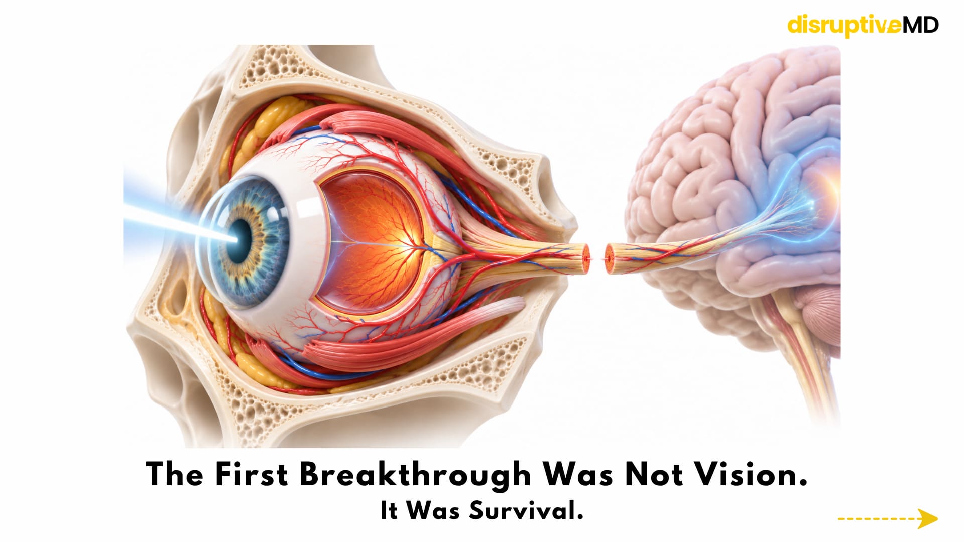
- A whole-eye transplant involves far more than moving the visible eyeball from one person to another.
- At one year, retinal and choroidal perfusion were still present.
- Yet there was no light perception in the transplanted eye.
THE FIRST BREAKTHROUGH WAS NOT VISION. IT WAS SURVIVAL.
The 2023 operation was extraordinary partly because of what had to remain intact.
A whole-eye transplant involves far more than moving the visible eyeball from one person to another. The globe must be transplanted together with critical vascular, neural, muscular, and orbital structures.
The donor eye needs an arterial blood supply.
Its venous drainage must work.
The retina cannot remain without oxygen for too long.
The optic nerve must be preserved as much as possible.
The extraocular muscles must eventually interact with the orbit.
And the transplanted tissue must survive the recipient’s immune system.
In the landmark human case, surgeons successfully revascularised the transplanted eye. At one year, retinal and choroidal perfusion were still present. Electroretinography — a test that measures electrical responses generated by the retina — detected residual responses to light.
Yet there was no light perception in the transplanted eye.
The 2026 follow-up strengthened the biological side of the story. The eye showed robust vascular perfusion and structural preservation over 12 months, with evidence that outer-retinal function had been maintained despite retinal nerve-fibre-layer loss and the severed optic nerve.
This distinction is crucial.
Keeping an eye alive is a transplantation problem.
Making that eye see is a neuroscience problem.
And the second may ultimately prove harder than the first.
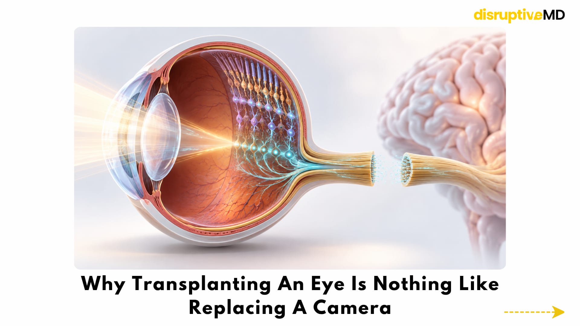
WHY TRANSPLANTING AN EYE IS NOTHING LIKE REPLACING A CAMERA
It is tempting to think of the eye as a biological camera.
The cornea and lens focus light.
The retina detects it.
The optic nerve acts like a cable carrying the information into the brain.
But the analogy quickly breaks down.
The retina is itself neural tissue containing multiple layers of specialised cells that begin processing visual information before it ever leaves the eye.
At the innermost output layer sit retinal ganglion cells, or RGCs.
Their long axons bundle together to form the optic nerve.
Those axons travel from the retina into the brain and ultimately communicate with visual processing centres.
This is where whole-eye transplantation runs into one of biology’s most difficult limitations.
The optic nerve is part of the central nervous system.
Unlike many peripheral nerves, damaged adult optic-nerve axons have very little natural regenerative capacity.
Simply placing two cut ends of an optic nerve together therefore does not recreate vision.
Thousands upon thousands of neural fibres would need to survive, regrow across the injury, travel through the recipient’s remaining visual pathway, reach appropriate brain targets and create functional connections.
And they would need to do this with enough organisation for the brain to interpret the signals as meaningful vision.
That is a radically different engineering problem from restoring blood flow.
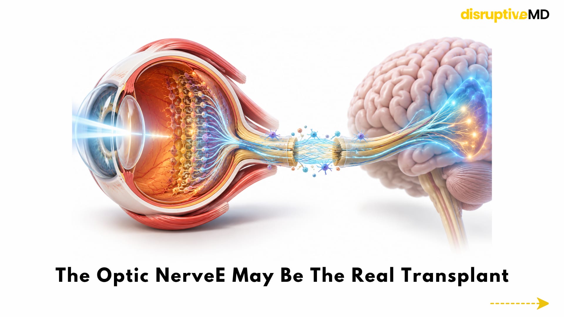
THE OPTIC NERVE MAY BE THE REAL TRANSPLANT
In one sense, whole-eye transplantation may eventually become less about the eyeball and more about the neural bridge between the eye and brain.
Current research is attacking that problem from several directions. Scientists are investigating ways to:
- Prevent retinal ganglion cells from dying after optic-nerve injury
- Switch mature neurons back into a growth-capable state
- Alter the inhibitory environment surrounding injured CNS axons
- Deliver neurotrophic and molecular growth signals
- Guide regenerating axons toward the correct brain targets
- Use gene-based approaches to increase regenerative capacity
- Replace lost retinal ganglion cells
- Create biomaterial scaffolds that physically guide axon growth
- Apply electrical or bioelectronic stimulation to encourage neural repair
Experimental research has already demonstrated that optic-nerve regeneration is biologically more modifiable than once assumed. But converting axon growth in laboratory models into accurate, durable human vision remains an enormous translational leap.
Growing a nerve is only part of the challenge.
The regenerated nerve has to connect to the right place.
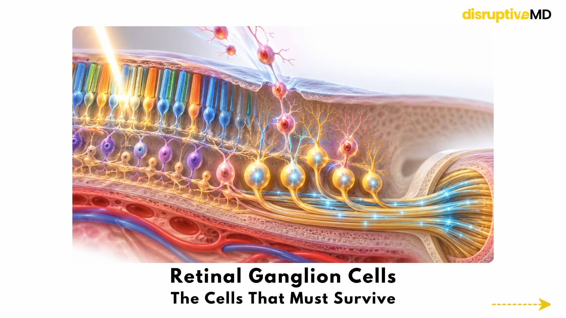
RETINAL GANGLION CELLS: THE CELLS THAT MUST SURVIVE
Retinal ganglion cells may become one of the most important cell types in the future of vision restoration.
They are the output neurons of the retina.
If they die, the photoreceptors may still react to light, but the eye loses its biological pathway for transmitting that information toward the brain.
This is one reason a transplanted eye can be structurally alive yet functionally blind.
Researchers are therefore studying not only how to regenerate existing RGC axons, but also whether damaged retinal ganglion cells themselves could eventually be replaced.
A particularly interesting advance came in 2026.
Experiments using laboratory-grown human retinal ganglion cells showed that the retina’s internal limiting membrane — a thin layer separating the vitreous from the neural retina — can act as an important physical barrier preventing transplanted RGCs from properly integrating into retinal tissue.
When that barrier was disrupted experimentally, transplanted human RGCs showed improved migration and structural integration in preclinical models.
This does not mean doctors can currently inject new ganglion cells and restore sight.
They cannot.
But it demonstrates the level at which the problem is now being investigated.
The future of eye transplantation could involve not only transplanting an organ, but reconstructing individual neuronal populations within that organ after transplantation.
BEFORE THE EYE CAN CONNECT TO THE BRAIN, IT HAS TO SURVIVE THE JOURNEY
Neural regeneration receives much of the attention, but there is another brutally simple problem:
The retina hates being without oxygen.
Between donor retrieval and surgical revascularisation, the transplanted eye can experience ischaemia — a period during which normal blood flow and oxygen delivery are interrupted.
Unlike some tissues, delicate neural structures such as the retina can be highly vulnerable to this injury.
The first human transplantation already demonstrated that a donor eye can survive the process.
But future sight-restoring transplantation will probably demand much better preservation.
The aim is not merely to preserve the shape of the globe.
It is to preserve the retinal neurons and optic-nerve machinery in the best possible biological condition before neural reconstruction even begins.
THE “ZERO WARM ISCHAEMIA” IDEA
One of the most interesting developments came in 2026.
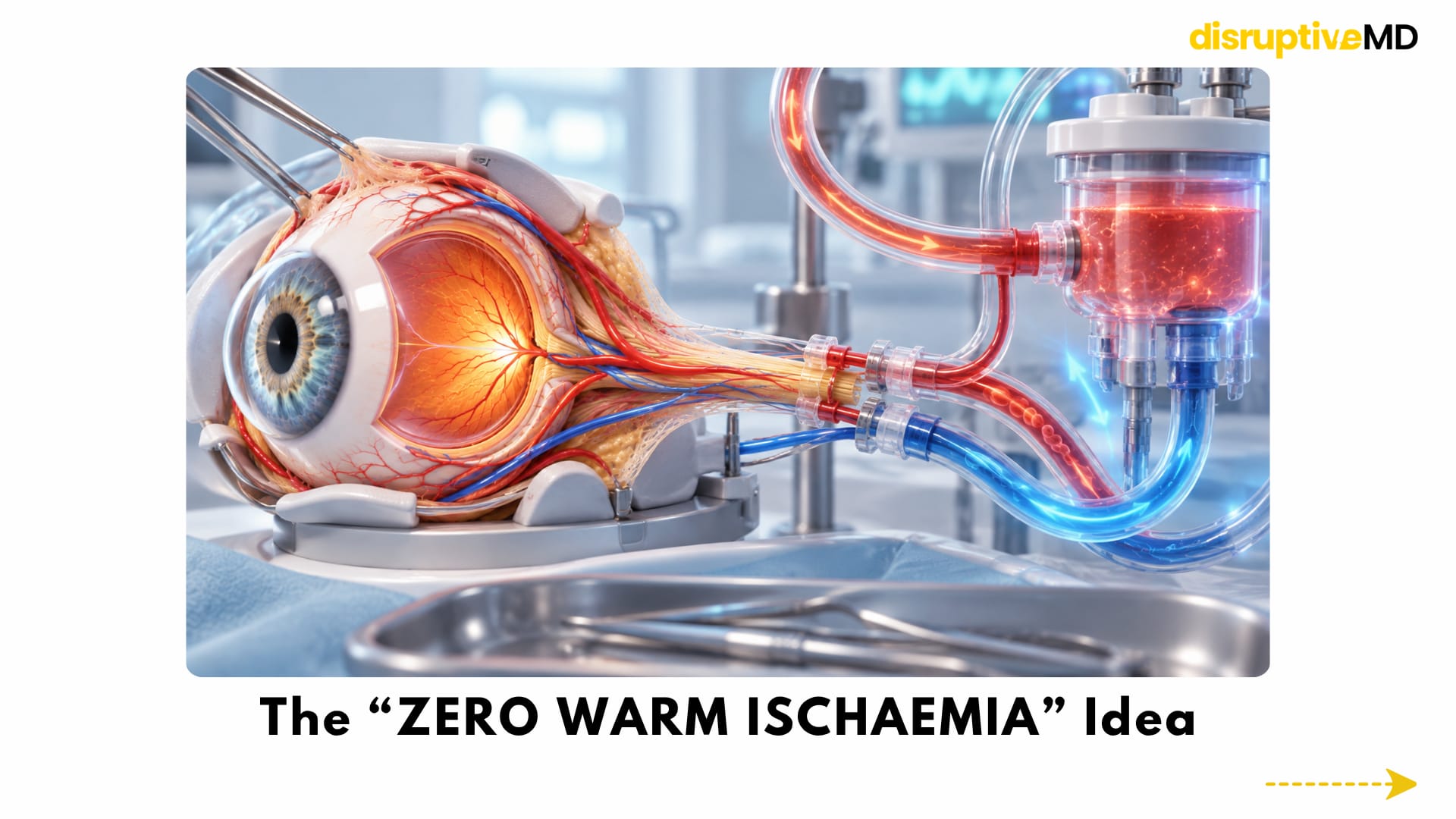
Researchers described a human cadaveric whole-eye transplantation technique designed to eliminate warm ischaemia time by maintaining continuous retrograde extracorporeal perfusion during donor-eye retrieval and simulated transplantation.
In two cadaveric procedures, continuous perfusion was maintained while the eye, optic nerve and associated structures were harvested and reconstructed, allowing the researchers to demonstrate the anatomical feasibility of a zero-warm-ischaemia transplantation protocol.
This was not a living-patient vision-restoration experiment.
It does not prove that the technique preserves functional sight.
But it represents an important conceptual shift.
Instead of accepting an unavoidable period during which the donor retina loses its circulation, future systems may attempt to keep the eye metabolically supported throughout much or all of the transplant process.
That could become enormously important if every surviving retinal ganglion cell and every preserved axon increases the biological starting point for later neural repair.
COULD A DONOR EYE BE KEPT ALIVE ON A MACHINE?
Another frontier is ex-vivo eye perfusion.
Modern transplantation increasingly uses machine-perfusion technologies to support donor organs outside the body.
A similar concept is now being explored for eyes: supplying a retrieved donor eye with controlled oxygenation, nutrients, protective compounds and circulation while clinicians assess whether the tissue remains suitable for transplantation.
Whole-eye transplantation research programs are actively developing donor-eye procurement and extracorporeal preservation technologies, including dedicated eye-perfusion systems.
Eventually, the donor eye may not simply be placed on ice and transported.
It could potentially become a monitored living graft outside the body — with clinicians assessing blood flow, retinal physiology, metabolism and neural integrity before implantation.
The preservation machine could become almost as important as the transplant operation itself.
MICROSURGERY HAS TO REBUILD THE PLUMBING
Even if the neuroscience were solved, the surgery would remain formidable.
The eye depends on extremely small vessels.
Whole-eye transplantation therefore sits at the intersection of ophthalmology, transplantation and high-precision reconstructive microsurgery.
Blood vessels must be reconnected with sufficient accuracy to rapidly restore perfusion without tension, thrombosis or inadequate flow.
The landmark human procedure demonstrated that this is surgically achievable.
Future procedures may become even more precise through advances in:
- Microsurgical instrumentation
- Supermicrosurgical techniques
- High-resolution intraoperative imaging
- Image-guided surgery
- Robotic tremor reduction
- Automated vessel measurement
- Perfusion monitoring
- Personalised surgical planning
The futuristic part is not necessarily replacing surgeons with robots.
It is giving surgeons increasingly precise control over structures measured in millimetres — or fractions of millimetres.
THE EYE ALSO HAS TO MOVE
Vision is not simply a retina detecting light.
A functional transplanted eye would have to become part of an entire oculomotor system.
The eye must point toward visual targets.
Extraocular muscles need appropriate attachment and control.
The eyelids have to open and close.
The ocular surface needs protection.
A stable tear film must be maintained.
The globe needs appropriate alignment within the orbit.
And if the other eye retains vision, the brain eventually has to coordinate information between both sides.
The 2023 transplant included reconstruction of the eye and surrounding facial/orbital structures, demonstrating that extraordinarily complex anatomical reconstruction is possible.
But functional vision would require much more than anatomical positioning.
A future seeing eye must behave as part of a coordinated sensory and motor network.
THEN COMES THE IMMUNE SYSTEM
A transplanted eye is foreign biological tissue.
That creates the same fundamental problem encountered in other forms of transplantation:
Rejection.
The first human whole-eye transplant occurred as part of a vascularised composite allotransplant and required systemic immunosuppression. During the reported first year, no acute rejection of the graft was documented.
That is encouraging.
But lifelong transplantation introduces another equation.
The potential benefit of restored vision would need to justify the risks of chronic immune suppression, including infection, metabolic complications, malignancy and drug toxicity.
Future transplantation therefore cannot focus only on nerve regeneration.
It will also need improved strategies for:
- Immune suppression
- Immune tolerance
- Inflammatory control
- Donor-recipient compatibility
- Rejection surveillance
- Graft monitoring
- Infection prevention
A biologically perfect optic nerve connection would mean little if the transplanted tissue could not survive long term.
A MAJOR PROGRAM IS NOW TRYING TO SOLVE THE WHOLE SYSTEM
Perhaps the clearest sign that whole-eye transplantation has moved from speculative surgery into an organised scientific field is the creation of the Transplantation of Human Eye Allografts — THEA — program.
The program is built around three major technical areas:
- Donor-eye retrieval and preservation
Keep the retina, optic nerve and other critical structures alive from donor retrieval until transplantation.
- Optic-nerve repair and regeneration
Enable retinal ganglion cell axons to survive, regrow and reconnect the transplanted eye with the brain.
- Surgery, postoperative care and functional assessment
Develop the transplantation techniques and technologies needed to determine whether the reconstructed eye is truly functioning.
The scale of the research matters because no single technology will make whole-eye transplantation work.
A perfect surgical transplant with a dead optic nerve will not restore sight.
Perfect nerve regeneration with a severely ischaemic retina will not restore sight.
A healthy retina and regenerated nerve will still fail if the immune system destroys the graft.
Whole-eye transplantation is therefore a systems problem.
It requires multiple technologies to mature together.
BIOELECTRONICS MAY BECOME PART OF THE SOLUTION
The most futuristic possibility is that the final solution may not be purely biological.
Some current whole-eye transplantation research programs explicitly include bioelectronic approaches to nerve regeneration.
That opens a provocative possibility.
What if a future transplanted eye cannot reconnect to the brain using biology alone?
Could technology help bridge the gap?
Possible future architectures could combine transplantation with:
- Retinal stimulation
- Optic-nerve interfaces
- Cortical visual prostheses
- Electrical-field stimulation
- Implantable neural interfaces
- Bioelectronic guidance systems
- Adaptive signal processing
- AI-assisted decoding of neural activity
None of these combinations has restored vision after a human whole-eye transplant.
They remain future concepts.
But whole-eye transplantation may eventually evolve into something more complex than conventional organ replacement:
A hybrid biological-neural interface in which surgery restores the organ, regenerative medicine rebuilds the pathway and electronics help reconnect information flow.
COULD THE BRAIN LEARN TO USE A TRANSPLANTED EYE?
Even successful nerve regeneration may not complete the job.
Ultimately, sight happens in the brain.
Retinal signals must reach central visual structures.
Those signals must preserve enough spatial and temporal organisation for downstream neural networks to interpret them.
And after a long period without visual input, the brain may itself have changed.
Neuroplasticity could therefore become another part of rehabilitation.
A future whole-eye transplantation program might not end when the surgical wounds heal.
It could require prolonged:
- Visual rehabilitation
- Neurostimulation
- Eye-movement retraining
- Perceptual learning
- Neural-interface calibration
- Functional imaging
- Adaptive visual training
The patient may not simply receive a new eye.
The brain may have to learn how to see through it.
WHO MIGHT RECEIVE THE FIRST FUNCTIONAL EYE TRANSPLANTS?
If functional whole-eye transplantation becomes possible, it is unlikely to immediately become a universal treatment for blindness.
Blindness is not one disease.
Different conditions destroy different components of the visual system.
If the recipient’s visual cortex or large portions of the central visual pathway are irreversibly damaged, transplanting a new eye may offer little advantage.
Early functional transplantation would therefore probably require extremely careful candidate selection.
One plausible initial population could include selected people with catastrophic traumatic eye loss in whom central visual pathways remain relatively preserved.
That is fundamentally different from assuming that whole-eye transplantation could immediately replace treatment for every form of glaucoma, macular degeneration, diabetic retinal disease or congenital blindness.
The technology will need to prove where the biological bottleneck lies for each patient.
WHAT IS REAL TODAY
Whole-eye transplantation is no longer entirely theoretical.
A complete human donor eye has been transplanted.
The transplanted globe can survive.
Blood flow can be successfully re-established.
Retinal tissue can remain structurally preserved for an extended period.
Residual retinal electrical responses to light can persist.
Microsurgical optic-nerve coaptation is anatomically possible.
Research into donor-eye machine preservation is active.
A zero-warm-ischaemia surgical strategy has been demonstrated in a human cadaveric model.
Human retinal ganglion-cell transplantation research is advancing.
Major research programs are directly targeting optic-nerve regeneration and functional eye transplantation.
These are genuine advances.
But they should not be confused with restored sight.
WHAT IS NOT REAL YET
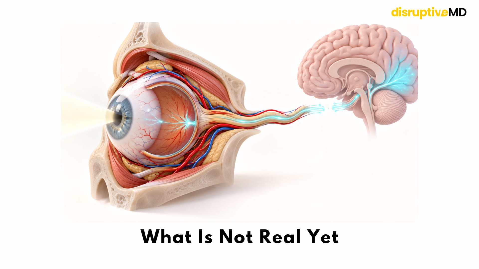
There has not yet been a human whole-eye transplant that restores functional vision.
Reliable regeneration of a severed human optic nerve has not been demonstrated clinically.
Transplanted retinal ganglion cells cannot yet be routinely integrated into the human retina to reconstruct a functional visual pathway.
There is no routine whole-eye transplant operation for blindness.
Long-term immune tolerance has not been solved.
Perfect donor-eye preservation has not been achieved.
And no clinical system currently reconnects an entire transplanted retina to the brain with normal visual processing.
That distinction matters enormously.
Whole-eye transplantation has crossed the boundary from impossible surgery to biological feasibility.
It has not yet crossed the boundary from biological feasibility to restored sight.
THE BIGGER STORY IS NOT REALLY ABOUT THE EYE
Whole-eye transplantation sits at an extraordinary intersection.
It brings together:
Transplantation
Ophthalmology
Microsurgery
Neuroscience
Stem-cell biology
Regenerative medicine
Gene therapy
Biomaterials
Bioelectronics
Neural engineering
and potentially AI-assisted neurotechnology.
That makes the field important even if routine eye transplantation remains years away.
If scientists learn how to make retinal ganglion cells regenerate long axons through an injured optic nerve and reconnect with the brain, the implications could extend far beyond blindness.
The optic nerve is part of the central nervous system.
Learning how to rebuild it could teach us something much bigger:
How to reconnect damaged neural tissue to the brain.
That is why the future of whole-eye transplantation may ultimately become a story about spinal cord repair, brain-machine interfaces and central nervous system regeneration as much as ophthalmology.
THE FIRST BREAKTHROUGH WAS KEEPING THE EYE ALIVE
For most of medical history, the question was:
Can an entire human eye even be transplanted?
We now have an early answer.
Yes. an entire donor eye can be surgically transplanted and kept biologically viable.
The question has changed.
Now it is:
Can the retina survive well enough?
Can retinal ganglion cells be protected or replaced?
Can millions of injured axons regenerate?
Can those axons find the correct targets inside the brain?
Can the immune system tolerate the graft?
And can all of those technologies work together in one person?
If the answer eventually becomes yes, whole-eye transplantation will represent something far more profound than another organ transplant.
It would mean medicine had learned to transplant not merely tissue —
but a sensory connection to the brain.
The first breakthrough was keeping the eye alive.
The next breakthrough would be making it see.
FACT BASE
Human feasibility: The first human whole-eye transplant was performed in May 2023 together with a partial-face transplant. The graft was successfully revascularised and remained viable. At one year, retinal and choroidal perfusion persisted, and electrophysiological testing showed residual retinal responses to light, but the transplanted eye had no light perception.
2026 follow-up: A 2026 report examining the first human whole-eye allotransplant documented robust vascular perfusion and structural preservation over 12 months. Outer-retinal electrophysiological function was maintained despite retinal nerve-fibre-layer loss and optic-nerve transection. Functional vision was not restored.
Optic-nerve barrier: The optic nerve consists primarily of retinal ganglion-cell axons and is part of the central nervous system. Mature mammalian optic-nerve axons have limited spontaneous regenerative capacity after injury, making functional reconnection one of the principal barriers to sight restoration.
Retinal ganglion-cell replacement: In 2026, preclinical research showed that disruption of the retinal internal limiting membrane could improve the migration and integration of transplanted laboratory-grown human retinal ganglion cells. The work advances RGC-replacement strategies but has not demonstrated restored human vision.
Donor-eye preservation: A 2026 cadaveric feasibility study demonstrated a whole-eye transplantation protocol using continuous retrograde extracorporeal perfusion to achieve zero warm ischaemia during simulated transplantation. This represents an anatomical and preservation advance, not a clinical demonstration of restored sight.
Machine perfusion: Dedicated research programs are developing extracorporeal systems intended to preserve donor-eye viability between retrieval and implantation. These technologies remain developmental rather than routine clinical transplantation tools.
Whole-eye transplantation research: The THEA program is organised around three core technical problems: donor-eye retrieval and preservation, optic-nerve repair and regeneration, and transplant surgery/postoperative functional assessment. Current funded work includes stem-cell, scaffold, regenerative and bioelectronic approaches.
Current clinical status: As of August 2026, whole-eye transplantation has demonstrated human surgical and biological feasibility, but functional vision restoration after human whole-eye transplantation has not been achieved.
REFERENCES
- Ceradini DJ, Tran DL, Dedania VS, et al. Combined Whole Eye and Face Transplant: Microsurgical Strategy and 1-Year Clinical Course. JAMA. 2024;332(18):1551–1558. DOI: 10.1001/jama.2024.12601.
- Dedania VS, Shah AR, Chinta SR, et al. Monitoring outcomes of the first human whole eye allotransplant. Documenta Ophthalmologica. 2026;152(2):235–246. DOI: 10.1007/s10633-026-10083-0.
- Li Y, et al. Emerging therapeutic strategies for optic nerve regeneration. Trends in Pharmacological Sciences. DOI: 10.1016/j.tips.2024.11.008.
- Gokoffski KK, Washington KM, Chuck RS. Clinical and Scientific Considerations for Whole Eye Transplantation: An Ophthalmologist’s Perspective. Translational Vision Science & Technology. 2025;14(2):13. DOI: 10.1167/tvst.14.2.13.
- Aguzzi EA, Mary S, Mowlavi Vardanjani M, et al. The internal limiting basement membrane inhibits functional engraftment of transplanted human retinal ganglion cells in vivo. Science Translational Medicine. 2026;18(847):eadr1062. DOI: 10.1126/scitranslmed.adr1062.
- Wang AC, Mousavi Motlagh YS, Amiri I, et al. Zero Warm Ischemia Time in Whole-Eye Transplantation through Retrograde Extracorporeal Perfusion. Ophthalmology Science. 2026;6(6):101183. DOI: 10.1016/j.xops.2026.101183.
- Wei L, Yan W, Zhang K, et al. Allogeneic whole-eye transplantation: advancements, challenges, and future directions in vision restoration. Frontiers in Medicine. 2025;12:1691259. DOI: 10.3389/fmed.2025.1691259.
- Transplantation of Human Eye Allografts — THEA. Program describing research priorities in donor-eye preservation, optic-nerve regeneration, transplantation and functional assessment.