
Introduction
Medical imaging has transitioned from seeing anatomy to helping doctors discover disease early, delivering more objective quantification, guiding treatment selection and enabling more precise monitoring of response. CT, MRI, PET, ultrasound and X-ray are still the fundamental diagnostic tools, but the next wave of innovation is transforming how pictures are captured, reconstructed, interpreted and related to treatment decisions. The most significant breakthroughs are not particular machines, but technology that change where imaging is done, what biological information it gives, how quickly results get to doctors and how reliably imaging can guide care. Among the most promising uses so far are PSMA PET for prostate cancer and several AI imaging technologies that are now FDA-approved. Additional fields such as portable MRI and broader implementation of AI require more real-world validation.
Medical Imaging Is Moving From Static Pictures to Actionable Data
The key issue in medical imaging is not if a scanner can make a clear picture, but if the picture can answer the clinical query swiftly, securely, equitably and precisely. Conventional imaging is generally reliant on costly infrastructure, experienced operators, professional interpretation and patient transfer to radiology departments, which can delay diagnosis in emergency care, rural settings, critical care units and under-resourced health systems. This is crucial as the decision to image typically affects whether a patient will have surgery, thrombolysis for stroke, cancer staging, radiation planning or targeted therapy.
The top five disruptive advances in medical imaging are artificial intelligence-assisted imaging, photon-counting CT, portable low-field MRI, targeted molecular imaging and theranostics, and handheld or AI-guided point-of-care ultrasound . They each hit a different area of the imaging path. AI is transforming image analysis and our work. Photon-counting CT is transforming X-ray detection. Portable MRI is transforming the location of MRI. Molecular imaging is transforming our view of disease biology. Point-of-care ultrasound is transforming who can obtain clinically valuable images at the bedside.


- AI-assisted imaging uses machine learning to support image reconstruction, organ segmentation, abnormality detection, triage, and quantitative assessment.
- Imaging is one of the strongest early areas for medical AI because scans generate large amounts of digital data that can be measured and prioritized.
- AI can help both analyse existing images and generate better imaging data, but safety, validation, and workflow risks remain important.
Top 5 innovations explained
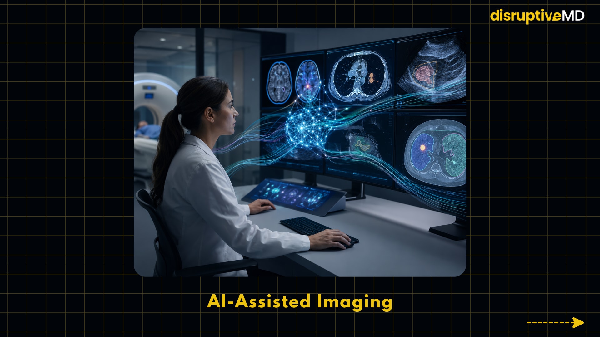
1. AI-assisted imaging: Images to quantifiable signals
Artificial intelligence-assisted imaging employs machine learning, software taught on data to learn to recognize patterns, to help with tasks such as image reconstruction, organ segmentation, identification of abnormalities, triage and quantitative assessment. This may include spotting a potential cerebral haemorrhage on CT in radiology, heart function on echo, denoising MRI, or triaging urgent studies for review. A 2025 assessment of 1,016 FDA authorizations of AI/ML-enabled medical devices found that 84.4% of unique AI devices used images as their core input, with the majority of image-based products examined by the radiology panel, demonstrating how deeply medical AI has entered imaging first. This is crucial since imaging creates a lot of digital data and well-validated AI can assist physicians manage workload, decrease repeated measuring activities and find time-sensitive findings faster.
The major technical difference is that AI can help with both “analysis” and “generation” of imaging data. Analysis involves the interpretation or measurement of existing images. e.g., lung nodule detection, ventricular volume calculation. Generation means helping to produce better imaging data for example by denoising, enhancing images, guiding acquisition or reconstructing. The FDA believes that AI-enabled medical devices need to be examined for safety and effectiveness, but that traditional medical device regulation was not created for adaptive AI systems that may evolve over time. This is crucial since AI in imaging is not one technology but a family of tools, with varying dangers depending on whether the program is merely measuring a structure, prioritizing a scan or influencing a diagnostic decision.
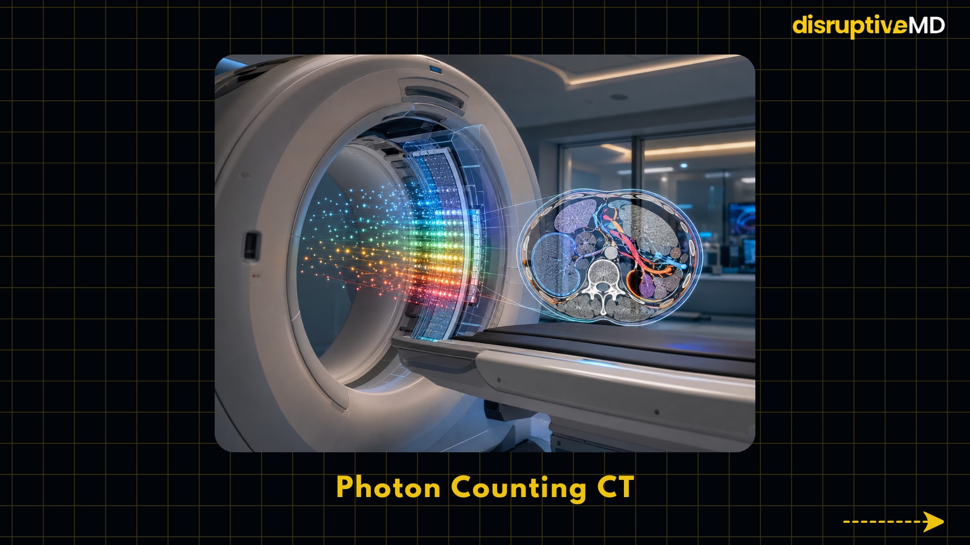
2. Photon counting CT: Better CT with more information from each X-ray photon
Photon-counting computed tomography, or photon-counting CT, or PCCT, modifies the detector in the CT scanner. In conventional CT detectors, many X-ray photons deposit their complete energy at the same moment, but photon-counting detectors count individual photons and sort them by their energy. This allows the scanner to obtain additional information regarding tissue composition and contrast material. According to a 2024 NCBI review, PCCT is intended to offer improved resolution, less radiation exposure, more tissue distinction and reduced electronic noise compared to traditional CT. This is of importance as CT is one of the most often employed imaging modalities in emergency care, cancer, trauma, cardiovascular disease, lung screening and surgical planning.
Photon-counting CT is particularly essential since it improves the physics of image production, not just add-on software after the scan. The FDA 510(k) documentation for a photon counting CT system describes the device as a computed tomography X-ray system with photon counting technology detectors that produces DICOM CT images for use by trained staff to aid in diagnosis, treatment and interventions. It’s a promise of not only nicer photos, but improved vision of small structures, metal-adjacent anatomy, iodine contrast, vascular disease, lung detail and perhaps lower-dose studies for patients who need repeated imaging.
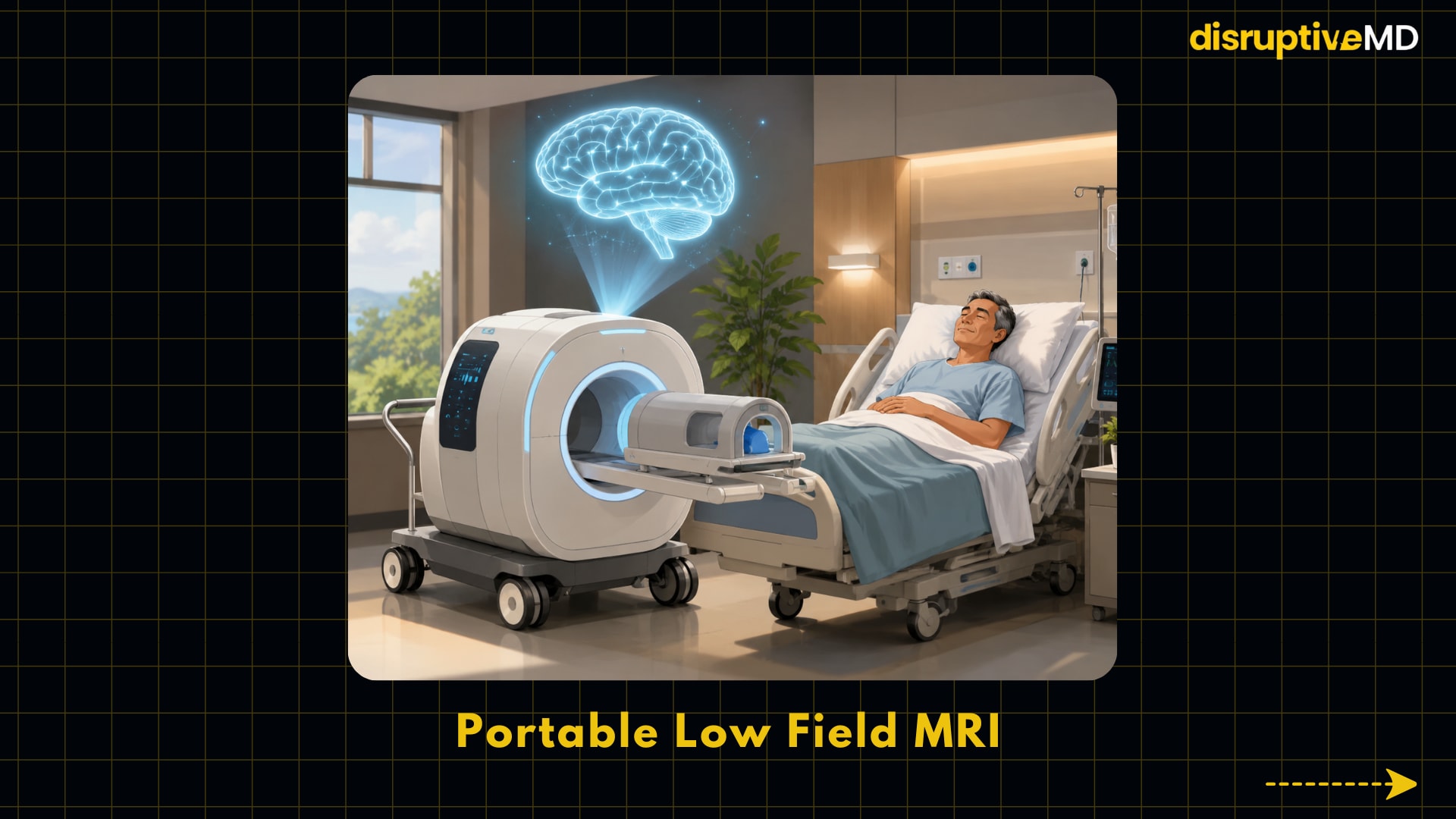
3. Portable low field MRI: Bringing MRI closer to the patient
Portable low field MRI uses compact hardware and contemporary image reconstruction and operates at a magnetic field that is orders of magnitude lower than the 1.5 T or 3 T fields used in standard hospital MRI scanners. “Low field” does not imply the same picture quality for all body parts or disorders, but rather that the scanner uses a lesser magnetic field that can reduce infrastructure requirements and imaging can be performed close to the patient’s bed side. Indeed, a 2025 assessment notes that current low-field systems benefit from improved algorithms for reconstruction, hardware miniaturization, and reduced infrastructure requirements, making them appropriate for point-of-care, remote, intraoperative, and limited-clinical settings. This is significant since MRI offers soft tissue contrast without ionizing radiation, and is difficult to reach with existing technology, especially for critically sick patients that cannot be safely moved, and in many parts of the world.
The most common usage is for brain imaging in intensive care and stroke assessment. A study on portable 0.064-tesla MRI for intracerebral hemorrhage published in Nature Communications included patients who had both conventional neuroimaging and portable MRI. Forty controls, 48 acute ischemic strokes, and 56 hemorrhages were assessed by neuroradiologists from 144 portable MRI scans. The study demonstrated a sensitivity of 80.4% for detection of intracerebral hemorrhage and a specificity of 96.6% for detection of blood-negative instances with no adverse effects during bedside scanning. This is essential because portable MRI may not replace comprehensive diagnostic MRI but rather could deliver neuroimaging to locations where transferring an unstable patient to a scanner would be risky or impractical.
4. Molecular imaging and theranostics: Viewing the illness biology, not anatomy
Targeted molecular imaging uses a radiotracer, a small amount of a radioactive chemical that binds to a biological target related to disease. PSMA PET is the best example right now . Prostate-specific membrane antigen (PSMA) is a protein that is typically overexpressed on prostate cancer cells. A PSMA PET scan involves injecting a radioactive chemical that binds to PSMA positive lesions, allowing a PET scanner to highlight areas where cancer is likely to be present. According to the National Cancer Institute, PSMA PET is an imaging technique that can aid to locate prostate cancer cells in the body and can help diagnosis recurrent or metastatic illness and plan treatment. This is significant since typically decisions about cancer treatment depend on whether there is genuinely localized, regional or metastatic disease.
The best clinical evidence is in prostate cancer stage The proPSMA randomised trial randomised 302 men with high-risk prostate cancer to either conventional imaging (CT and bone scan) or gallium-68 PSMA-11 PET-CT. PSMA PET-CT was 92% accurate compared with 65% for conventional imaging, generated fewer equivocal findings and resulted in more management changes. In the 2025 pocket guideline of the European Association of Urology, PSMA-PET/CT is strongly recommended for metastatic screening in high-risk localized or locally progressed prostate cancer, if available. This is of importance as proper staging may prevent undertreatment of metastatic disease and overtreatment of local treatment in some patients.
Theranostics brings together therapy and diagnostics and using imaging to identify patients for a matched tailored treatment. The FDA says gallium Ga 68 gozetotide may be used as a PET imaging agent to locate patients who are candidates for lutetium Lu 177 vipivotide tetraxetan, a radioligand therapy directed against PSMA, in patients with PSMA-positive metastatic castration-resistant prostate cancer. The phase 3 VISION trial, in which 831 patients were randomized, showed median imaging-based progression-free survival improving from 3.4 to 8.7 months and median overall survival improving from 11.3 to 15.3 months, but the radioligand therapy was associated with more grade 3 or higher adverse events. This is essential as imaging is no longer merely diagnostic in this approach, it is becoming a gatekeeper to precise treatment.
5. Point-of-care ultrasound: Portable, AI-enhanced bedside imaging
Point-of-care ultrasound (POCUS) is the use of portable ultrasonography at the bedside to answer a specific clinical question: is the heart pumping badly, is there fluid in the belly, is the pregnancy viable, can a procedure be performed more safely? Ultrasound imaging may be performed in real time, is more portable than CT or MRI and does not require ionizing radiation. This is crucial because many urgent clinical decisions are made outside the radiography suite in EDs, ICUs, ambulances, rural clinics, operating rooms, primary care settings.
The disruptive element is not just miniaturization, but also the AI-guided acquisition and the automated measurement. In a prospective multicentre study in npj Digital Medicine, 424 studies with analysable results compared focused cardiac ultrasonography with AI-assisted left ventricular ejection fraction quantification with formal transthoracic echocardiography. The AI-assisted bedside approach has an overall intraclass correlation coefficient of 0.904, an AUC of 0.98 for abnormal ejection fraction <50% detection, and a sensitivity of 92.8% and specificity of 92.3%. This is important as left ventricular ejection fraction is an important measure of the amount of blood pumped by the heart with each beat and reliable bedside estimation could allow for faster decision making in heart failure, shock and perioperative settings when formal echocardiography is delayed.
Evidence and Real-World Meaning
The evidence base for AI-assisted imaging is large yet uneven. The FDA maintains a public database of AI-enabled medical devices that have been granted marketing clearance to be used in the United States, and recent analysis suggest imaging is the most common domain of this regulatory landscape. Regulatory approval, however, does not ensure that all AI tools will improve patient outcomes in real-world practice. In a 2025 cross-sectional analysis published in JAMA Network Open of 903 FDA-approved AI-enabled devices, clinical performance studies were reported for 505 devices (55.9%) and clearly stated for 218 devices (24.1%) that no clinical performance studies were performed. Only 41 of the clinical investigations were prospective, and 12 were randomized. This is significant since an AI model that has good performance on the development dataset may not generalize well to other scanner kinds, hospitals, patient groups, illness prevalence, and workflow conditions.
Photon counting CT offers compelling engineering reasoning and increasing clinical use, but the entire population level impact is still being determined. Technical reviews strongly support the technical advantages of physics (reduction of electronic noise, improved spatial resolution, improved spectral information, and potential dose reduction). The FDA device summaries confirm clinical use of CT systems incorporating photon-counting detector technology. The real-world meaning is most likely to have clinical implications in areas where small structures, material differentiation, low dose imaging or contrast reduction are clinically important such as pediatric imaging, coronary CT angiography, lung disease, kidney stone characterization, gout imaging and metal implant evaluation.
Promising observational data are available for portable low-field MRI, especially for bedside neuroimaging, although this technology still remains a targeted approach, not a universal replacement for conventional MRI. The 2021 intracerebral hemorrhage research indicated feasibility and relevant diagnostic performance although 80.4% sensitivity means some hemorrhages were missed and picture quality is greatly reliant on anatomy, sequence design, disease type and scanner field strength. Thus, the real-world implication is widening of access: portable MRI may provide triage, monitoring and repeat imaging when conventional MRI is inaccessible or risky to access, but should be paired with specialist interpretation and well-defined protocols.
Among those mentioned here, targeted molecular imaging has some of the strongest clinical data, notably PSMA PET for prostate cancer. The proPSMA trial showed increased staging accuracy and fewer equivocal results compared with CT and bone scan and professional guidelines now advocate PSMA PET/CT in select prostate cancer staging scenarios where applicable. Additional theranostic application adds to the evidence base as imaging can identify PSMA positive illness prior to PSMA focused radioligand therapy and randomized trial data demonstrates survival benefit in selected patients with advanced prostate cancer. In practical terms, that implies imaging is starting to be used as a biologic selection tool, not merely a map of where the tumor is.
The AI-guided portable ultrasound is attractive in practice but requires further proof across body systems, operators and contexts. The cardiac ejection fraction study reveals that AI-assisted focused ultrasound may mimic formal echocardiography for a specific measurement in a multicentre cohort, but that does not mean that every bedside ultrasound inquiry can be securely automated. The real-world implication is speed and access. Well-validated AI-guided POCUS can help non-specialist clinicians obtain interpretable pictures and quantitative data, especially in resource-limited situations with restricted access to sonographers and radiologists.
Limitations, Risks, and Unanswered Questions
The biggest danger with these advancements is the assumption that technical excellence will automatically mean better outcomes for patients. AI tools might be subject to dataset shift, i.e. the performance can fluctuate when a model is confronted with patients, scanners, illness patterns or procedures different from those used for training and validation . Similar gaps exist in public reporting of many AI-enabled devices, with the 2025 JAMA Network Open analysis revealing missing or inadequate public information on clinical generalizability, including age- and sex-specific subgroup performance. This matters because if imaging methods are less reliable in underserved groups, they can exacerbate inequalities.
Questions of cost, availability, protocol standardization and evidence for photon counting CT remain. With greater imaging resolutions, more incidental findings, unexpected abnormalities that may not be clinically important, may be seen, and health systems will have to decide when PCCT offers enough added value to be implemented. The key question that remains open is not whether the detector technology is better in principle, but what clinical indications lead to better decisions, fewer repeat scans, less overall radiation, or better results.
Each clinical application of portable low-field MRI should be thoroughly validated. The lower magnetic field strength generally leads to a worse signal-to-noise ratio, so images may be less detailed, unless adjusted by the hardware and the reconstruction software. Portable MRI may increase access, but doctors need to know when bedside imaging is adequate and when traditional MRI or CT is still required. The key restriction is that early investigations are promising, but not adequate to establish portable MRI as a universal diagnostic option.
Molecular imaging and theranostics raise issues of access, cost, radiation logistics, isotope supply and suitable patient selection. PSMA targeted imaging in prostate cancer is well supported by evidence although positive uptake can be detected in a range of non-prostate disorders, and the interpretation of imaging still requires skilled specialists. PSMA-directed radioligand therapy also includes clinically meaningful toxicities such as myelosuppression and renal damage and FDA notifications highlight radiation exposure hazards. Precision imaging will only improve care if the right patients, tracers, treatment infrastructure and follow-up procedures are in place. This is crucial.
Point-of-care ultrasonography may enhance access to imaging but can also provide false reassurance if operators overestimate image quality or use automated metrics outside of defined indications. Ultrasound is operator dependent and the quality of pictures obtained is greatly dependent on probe position, patient anatomy and scanning expertise. AI guidance could alleviate this dependence, however the evidence so far is strongest for specialized tasks such as focused cardiac ejection fraction prediction, but not for other emergency or primary care ultrasonography use cases.
Conclusion
Medical imaging will likely become more quantitative, portable, biologically specific and integrated with treatment decisions in the future. Artificial intelligence may reduce repetitive tasks and facilitate faster triage, photon-counting CT may extract more information from X-ray imaging, portable MRI may bring advanced neuroimaging closer to the critically ill or underserved patient, molecular imaging may link diagnosis directly to targeted therapy and AI-guided ultrasound may extend bedside imaging beyond specialist departments. The realistic future is not one of replacing professionals with machines but of better tools to assist clinicians ask sharper questions, select imaging more wisely and respond sooner when the evidence is there to justify action. The most crucial metric of success will not be picture sharpness alone, but whether new technologies improve diagnostic confidence, patient outcomes, safety, access and health system efficiency.
Evidence Rating
The evidence is minimal or mixed. Several innovations are already allowed by authorities for some applications, including AI-enabled imaging devices, photon counting CT systems, PSMA-PET agents and PSMA-directed radioligand treatment. There is substantial guideline and clinical trial evidence for PSMA PET and PSMA theranostics in specific prostate cancer scenarios. Photon counting CT has good technical and early clinical support but needs additional data on broad implementation outcomes. Portable low-field MRI and AI-guided POCUS are clinically promising but are more hampered by indication-specific validation, workflow issues and generalizability limitations.
Educational Disclaimer
This material is for educational purposes only and does not substitute for professional medical advice, diagnosis, treatment or imaging interpretation. “Patients and caregivers should consult qualified healthcare professionals regarding medical imaging, diagnosis or therapy decisions.”
References
- U.S. Food and Drug Administration. Artificial Intelligence-Enabled Medical Devices.
- U.S. Food and Drug Administration. Artificial Intelligence in Software as a Medical Device.
- Singh, R., Bapna, M., Diab, A.R. et al. How AI is used in FDA-authorized medical devices: a taxonomy across 1,016 authorizations. npj Digit. Med. 8, 388 (2025). https://doi.org/10.1038/s41746-025-01800-1.
- Windecker D, Baj G, Shiri I, et al. Generalizability of FDA-Approved AI-Enabled Medical Devices for Clinical Use. JAMA Netw Open. 2025;8(4):e258052. doi:10.1001/jamanetworkopen.2025.8052.
- Lachance C, Horton J; Authors. Photon-Counting CT: High Resolution, Less Radiation: Emerging Health Technologies [Internet]. Ottawa (ON): Canadian Agency for Drugs and Technologies in Health; 2024 Feb. Available from: https://www.ncbi.nlm.nih.gov/books/NBK602525/.
- U.S. FDA 510(k) Summary. NAEOTOM Alpha photon-counting CT documentation.
- Kravchenko, D., Hagar, M.T., Vecsey-Nagy, M. et al. Low-field and portable MRI technology: advancements and innovations. Eur Radiol Exp 9, 103 (2025). https://doi.org/10.1186/s41747-025-00638-2.
- Mazurek, M.H., Cahn, B.A., Yuen, M.M. et al. Portable, bedside, low-field magnetic resonance imaging for evaluation of intracerebral hemorrhage. Nat Commun 12, 5119 (2021). https://doi.org/10.1038/s41467-021-25441-6.
- National Cancer Institute. PSMA PET scan definition.
- Michael S Hofman, Nathan Lawrentschuk, Roslyn J Francis, Colin Tang, Ian Vela, Paul Thomas, Natalie Rutherford, Jarad M Martin, Mark Frydenberg, Ramdave Shakher, Lih-Ming Wong, Kim Taubman, Sze Ting Lee, Edward Hsiao, Paul Roach, Michelle Nottage, Ian Kirkwood, Dickon Hayne, Emma Link, Petra Marusic, Anetta Matera, Alan Herschtal, Amir Iravani, Rodney J Hicks, Scott Williams, Declan G Murphy,Prostate-specific membrane antigen PET-CT in patients with high-risk prostate cancer before curative-intent surgery or radiotherapy (proPSMA): a prospective, randomised, multicentre study,The Lancet,Volume 395, Issue 10231,2020,Pages 1208-1216,ISSN 0140-6736,https://doi.org/10.1016/S0140-6736(20)30314-7.
- European Association of Urology. EAU-EANM-ESTRO-ESUR-ISUP-SIOG Prostate Cancer Pocket Guidelines. 2025.
- U.S. Food and Drug Administration. FDA approval of Pluvicto and Locametz. 2022.
- Sartor O, et al. Lutetium-177–PSMA-617 for Metastatic Castration-Resistant Prostate Cancer. New England Journal of Medicine. 2021.
- Sartor O, de Bono J, Chi KN, Fizazi K, Herrmann K, Rahbar K, Tagawa ST, Nordquist LT, Vaishampayan N, El-Haddad G, Park CH, Beer TM, Armour A, Pérez-Contreras WJ, DeSilvio M, Kpamegan E, Gericke G, Messmann RA, Morris MJ, Krause BJ; VISION Investigators. Lutetium-177-PSMA-617 for Metastatic Castration-Resistant Prostate Cancer. N Engl J Med. 2021 Sep 16;385(12):1091-1103. doi: 10.1056/NEJMoa2107322. Epub 2021 Jun 23. PMID: 34161051; PMCID: PMC8446332.
- Ferraz S, Coimbra M and Pedrosa J (2023) Assisted probe guidance in cardiac ultrasound: A review. Front. Cardiovasc. Med. 10:1056055. doi: 10.3389/fcvm.2023.1056055.