
What if one scan could search your entire body for disease before symptoms appear but also find problems that were never going to harm you?
The idea sounds like the ultimate expression of preventive medicine.
A person enters a scanner feeling completely healthy. Within a relatively short examination, large areas of the body are searched for hidden cancers, vascular abnormalities, organ disease, inflammation and structural changes.
The promise is powerful: Find disease before symptoms begin. Treat it before it spreads. Prevent a medical crisis before it happens.
In an age of artificial intelligence, genetic risk prediction, liquid biopsy, advanced imaging and continuous health monitoring, full-body scanning appears to offer a glimpse of medicine’s future.
But the reality is more complicated.
The human body is not a perfectly clean anatomical machine. Healthy people frequently have cysts, nodules, benign tumours, old injuries, anatomical variations and age-related changes. Most will never become dangerous.
The more closely medicine looks, the more abnormalities it finds.
That creates a difficult question: Does full-body scanning save lives? Or does it turn healthy people into patients?
The answer depends not only on what a scan can detect, but on whether detecting it improves the person’s outcome.
- Prevention reduces the probability that disease will develop.
- A scan searches for disease or abnormality that may already exist.
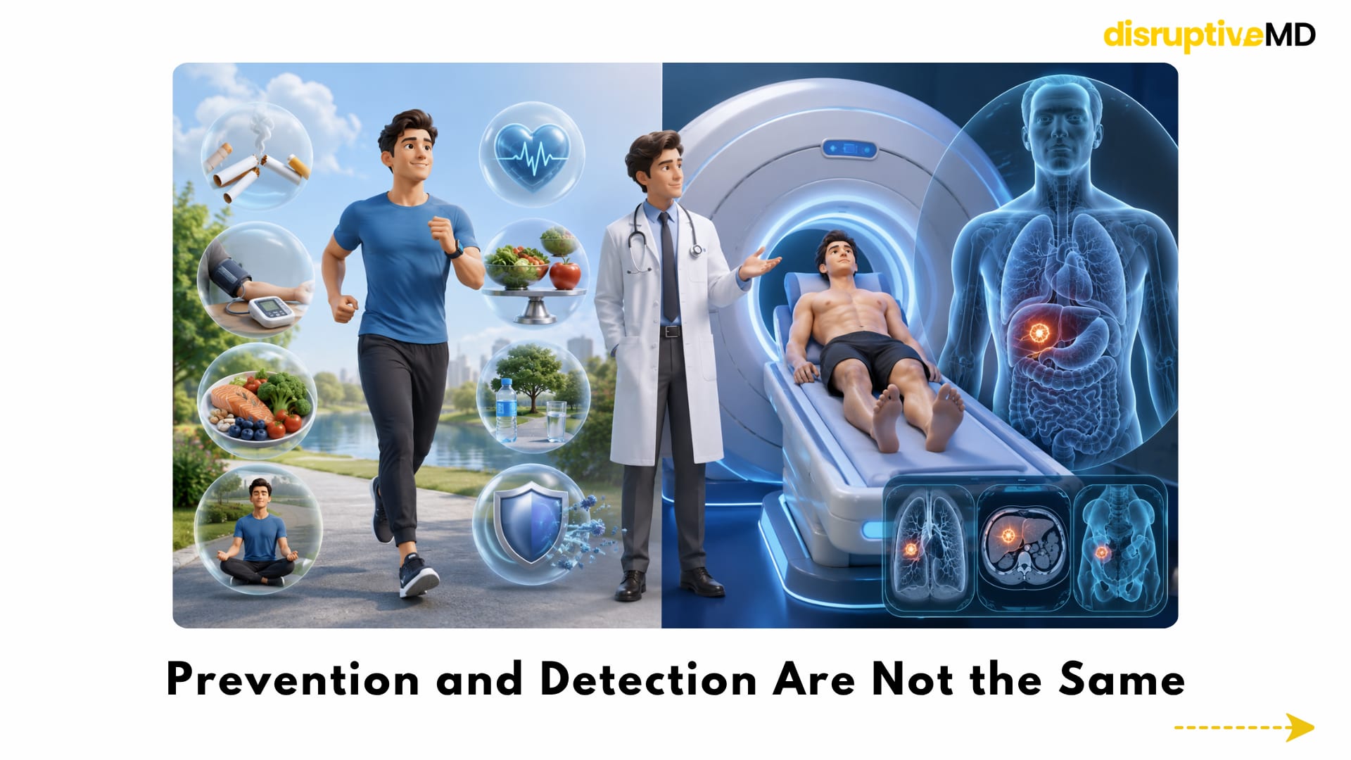
- Full-body scanning is a form of early detection rather than prevention itself.
Prevention and Detection Are Not the Same
Full-body scans are often marketed as preventive healthcare.
However, scanning is not prevention in the strict sense.
Prevention reduces the probability that disease will develop. Vaccination, smoking cessation, physical activity, metabolic health, blood-pressure control and reducing harmful exposures are forms of prevention.
A scan does something different.
It searches for disease or abnormality that may already exist.
That makes full-body scanning a form of early detection rather than prevention itself.
This distinction matters because earlier detection is not automatically beneficial.
Early detection is valuable when all of the following are true:
- the disease is important enough to cause future harm;
- the scan can identify it accurately;
- finding it earlier creates a meaningful treatment advantage;
- treatment at the detected stage improves outcomes;
- the benefits outweigh false positives, procedures, anxiety and cost.
A scan that finds more abnormalities has not necessarily created better healthcare.
The real test is whether the complete screening pathway reduces advanced disease, disability or death without causing greater harm elsewhere.
What Is a Full-Body Scan?
“Full-body scan” is not one single, standardised medical test.
It may refer to different imaging technologies, scan durations, anatomical coverage, imaging sequences and reporting methods.
Some examinations include the brain, neck, chest, abdomen and pelvis. Others may include the spine, bones, blood vessels or limbs. Some use contrast agents, while others do not.
The quality and purpose of the examination can therefore vary considerably.
A comprehensive scan designed for inherited cancer surveillance is not necessarily the same as a short commercial wellness scan.
Likewise, a full-body examination is not automatically equal to several dedicated organ-specific scans.
A broad scan must cover a very large anatomical area in a limited period. A dedicated examination can concentrate on one organ using sequences, contrast timing, positioning and image resolution specifically designed for that clinical question.
This creates an important trade-off: Full-body imaging offers breadth, but organ-specific imaging may offer greater depth.
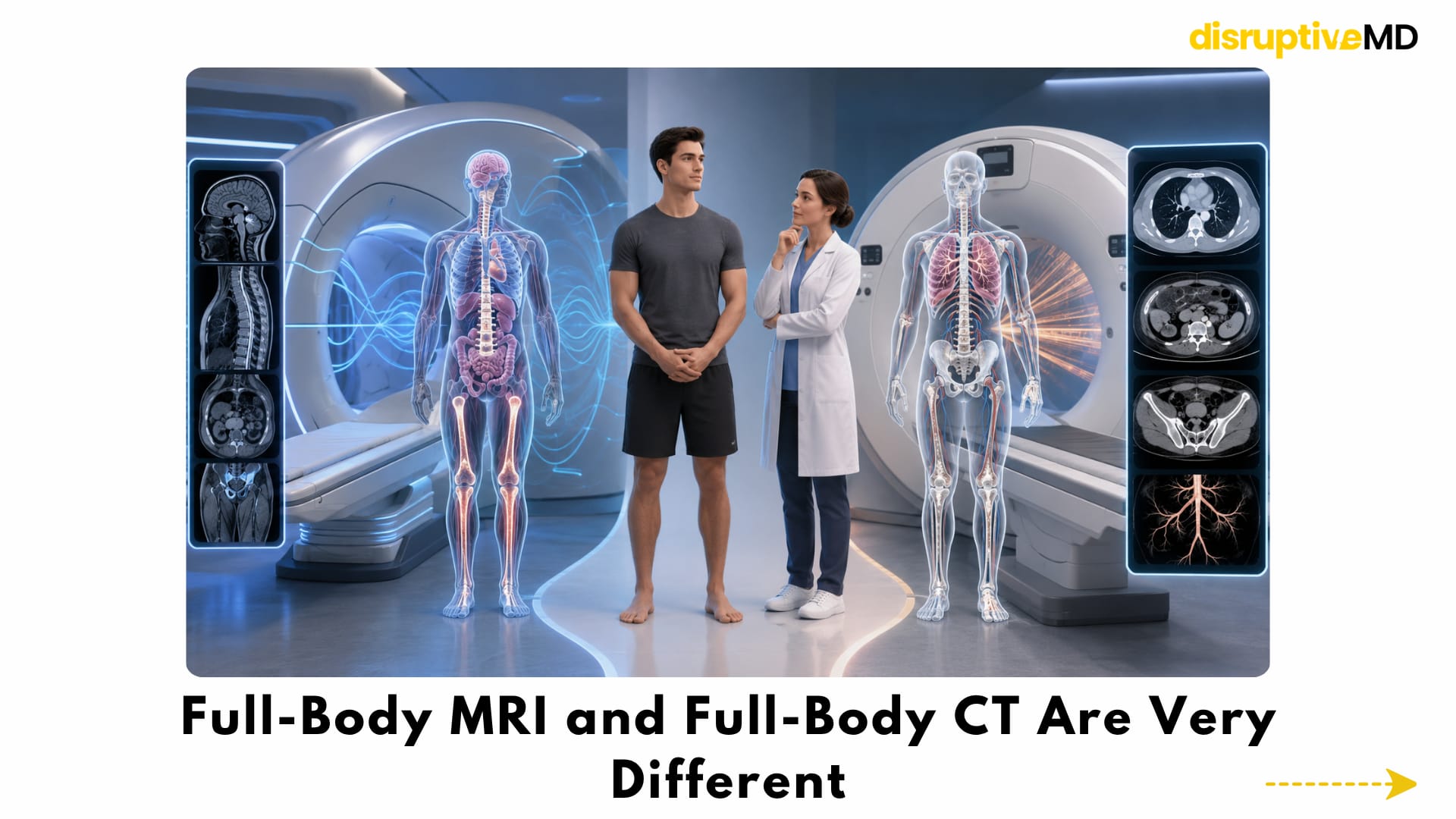
Full-Body MRI and Full-Body CT Are Very Different
The phrase “full-body scan” may describe MRI or CT, but these technologies are not interchangeable.
Full-body MRI
MRI uses magnetic fields and radiofrequency signals to create images. It does not expose the person to ionising radiation.
It is particularly useful for examining many soft tissues, organs, bone marrow and the central nervous system. Modern protocols may also use diffusion-weighted imaging, which assesses the movement of water molecules through tissue and can help identify areas with increased cellular density.
MRI is therefore attractive for repeated surveillance in carefully selected patients, particularly when radiation exposure should be minimised.
However, MRI also has limitations.
It may be less effective than dedicated imaging for certain small lung abnormalities, calcification, some thyroid lesions, parts of the gastrointestinal tract and several organ-specific cancers. Broad protocols can also differ substantially between centres.
A 2024 retrospective study involving 2,064 asymptomatic individuals found confirmed cancer in 1.2% of participants. However, supplementary examinations detected three cancers that had not been classified as highly suspicious on the whole-body MRI, illustrating that a broad MRI examination does not eliminate blind spots.
Full-body CT
CT creates detailed cross-sectional images using X-rays.
It is fast and can provide excellent information about the lungs, bones, blood vessels, calcification and many acute abnormalities.
The major difference is radiation.
A medically necessary CT can provide benefits that greatly outweigh its radiation risk. But the calculation changes when CT is used in healthy, low-risk individuals who may repeat the examination over time.
Radiation exposure is cumulative, and the estimated risk varies with age, sex, body region, scan protocol and total dose.
An analysis of elective full-body CT estimated that one examination in a 45-year-old adult could produce a small additional lifetime risk of cancer mortality, with repeated annual examinations increasing that estimated risk.
This does not mean CT should be feared when clinically needed.
It means that exposing healthy people to radiation requires a strong expectation of meaningful benefit.
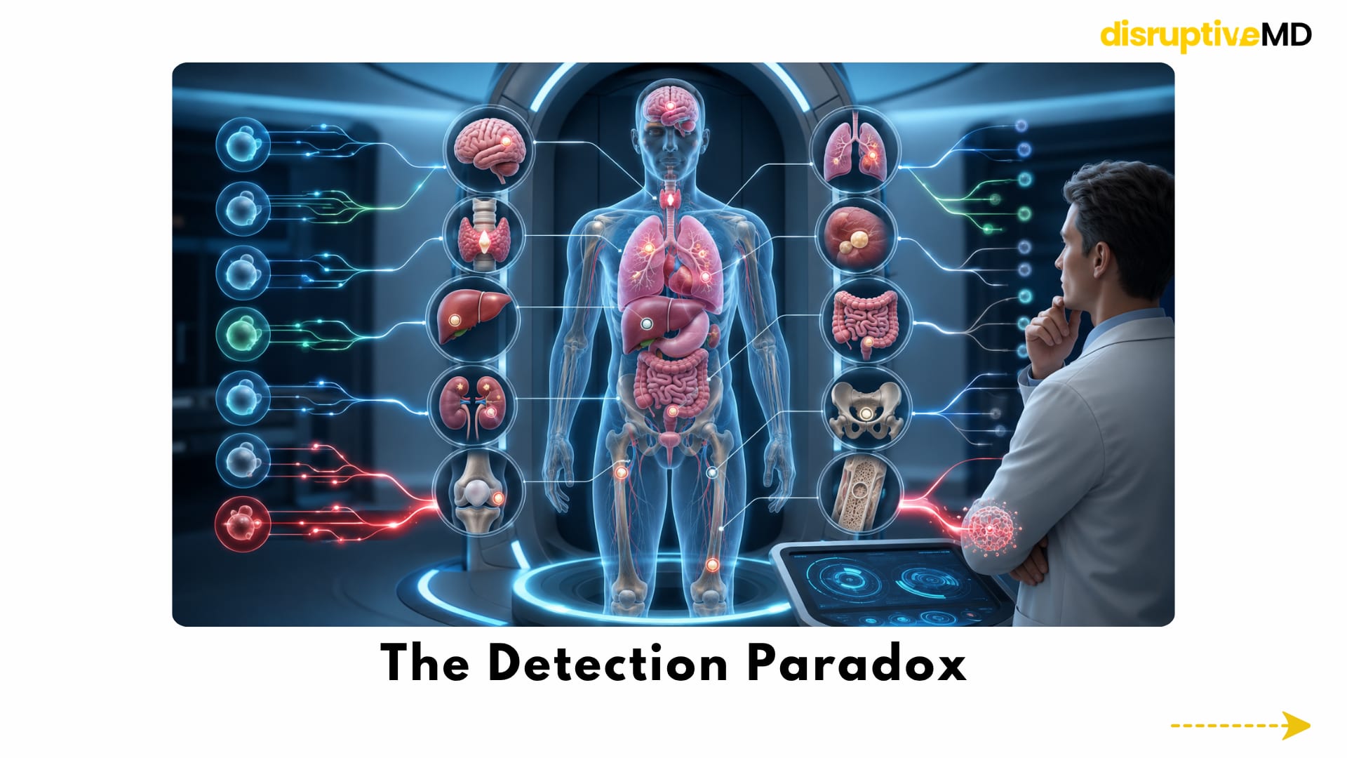
The Detection Paradox
Full-body imaging creates a paradox.
Its greatest strength is also its greatest weakness.
The technology can examine many organs at once.
But every additional organ creates another opportunity to find something uncertain.
Inside a large group of healthy people, scanners may identify:
- kidney cysts;
- liver cysts;
- thyroid nodules;
- lung nodules;
- adrenal masses;
- pancreatic cysts;
- benign tumours;
- vascular variations;
- spinal degeneration;
- joint changes;
- inflammation;
- old fractures or injuries;
- disc abnormalities;
- anatomical variants;
- small lesions of uncertain importance;
- occasionally, a serious hidden disease.
The central problem is not finding abnormalities.
It is deciding which abnormalities have the biological potential to become dangerous.
A scan shows structure and, in some cases, functional imaging characteristics. It does not always reveal how a lesion will behave over the next five, ten or twenty years.
A tiny abnormality may be:
- an early aggressive disease;
- a slow-growing disease;
- a harmless benign change;
- an imaging artefact;
- a finding that can never be fully classified without another test.
The image may be highly detailed while the future remains uncertain.
The Incidentaloma Problem
An incidentaloma is an unexpected abnormality discovered during imaging performed for another reason, or during broad screening when no specific disease was suspected.
Many incidental findings are harmless.
But once an abnormality is documented, it can be difficult to leave it alone.
The finding may begin a diagnostic chain reaction:
- A scan identifies a small abnormality.
- A dedicated scan is recommended.
- The second scan remains uncertain.
- Short-term surveillance is advised.
- A specialist consultation follows.
- A biopsy or procedure is considered.
- The patient spends months living with uncertainty.
The original scan may have taken less than an hour.
The consequences can continue for years.
A systematic review of whole-body MRI screening in asymptomatic people found that critical or indeterminate incidental findings were common, while many findings did not receive complete verification or long-term follow-up. The pooled estimate for critical or indeterminate findings was approximately 32%, although results varied greatly between studies and protocols.
Another large systematic review of brain and body MRI in apparently asymptomatic adults also found that potentially serious incidental findings were not rare and that their long-term consequences remained insufficiently understood.
This is why the value of full-body scanning cannot be judged only by the number of diseases detected.
Every positive or uncertain finding creates downstream consequences that must also be counted.
What Happens After an Incidental Finding?
The next step depends on the organ, size, appearance and estimated risk of the abnormality.
Possible follow-up may include:
- repeat MRI;
- ultrasound;
- CT;
- blood tests;
- endoscopy;
- specialist assessment;
- surveillance over several years;
- needle biopsy;
- surgical biopsy;
- removal of the abnormality.
Some of these actions are clearly justified.
Others may eventually prove unnecessary.
In a population-based whole-body MRI study, disclosure of tumour-related incidental findings increased the number of biopsies performed. Some biopsies identified malignancy, while many detected benign conditions.
This demonstrates the central difficulty.
Additional testing is not automatically wasteful because some important diseases will be found.
But a screening strategy must show that the benefit from those discoveries exceeds the cumulative harm experienced by everyone who undergoes unnecessary investigation.
False Positives
A false positive occurs when a scan suggests that a concerning disease may be present, but subsequent testing shows that it is not.
For example, an irregular mass may look suspicious on an initial broad scan. A dedicated scan, biopsy or surgical procedure may later show that it is benign.
The final diagnosis may be reassuring.
The journey to that diagnosis may still involve:
- intense anxiety;
- time away from work;
- specialist appointments;
- financial cost;
- additional radiation;
- contrast exposure;
- invasive procedures;
- complications;
- months of uncertainty.
The psychological impact should not be underestimated.
The person entered the scanner feeling healthy.
They may leave believing that cancer is present until further testing proves otherwise.
Even after a benign diagnosis, some people continue to worry that something was missed or that the lesion could change.
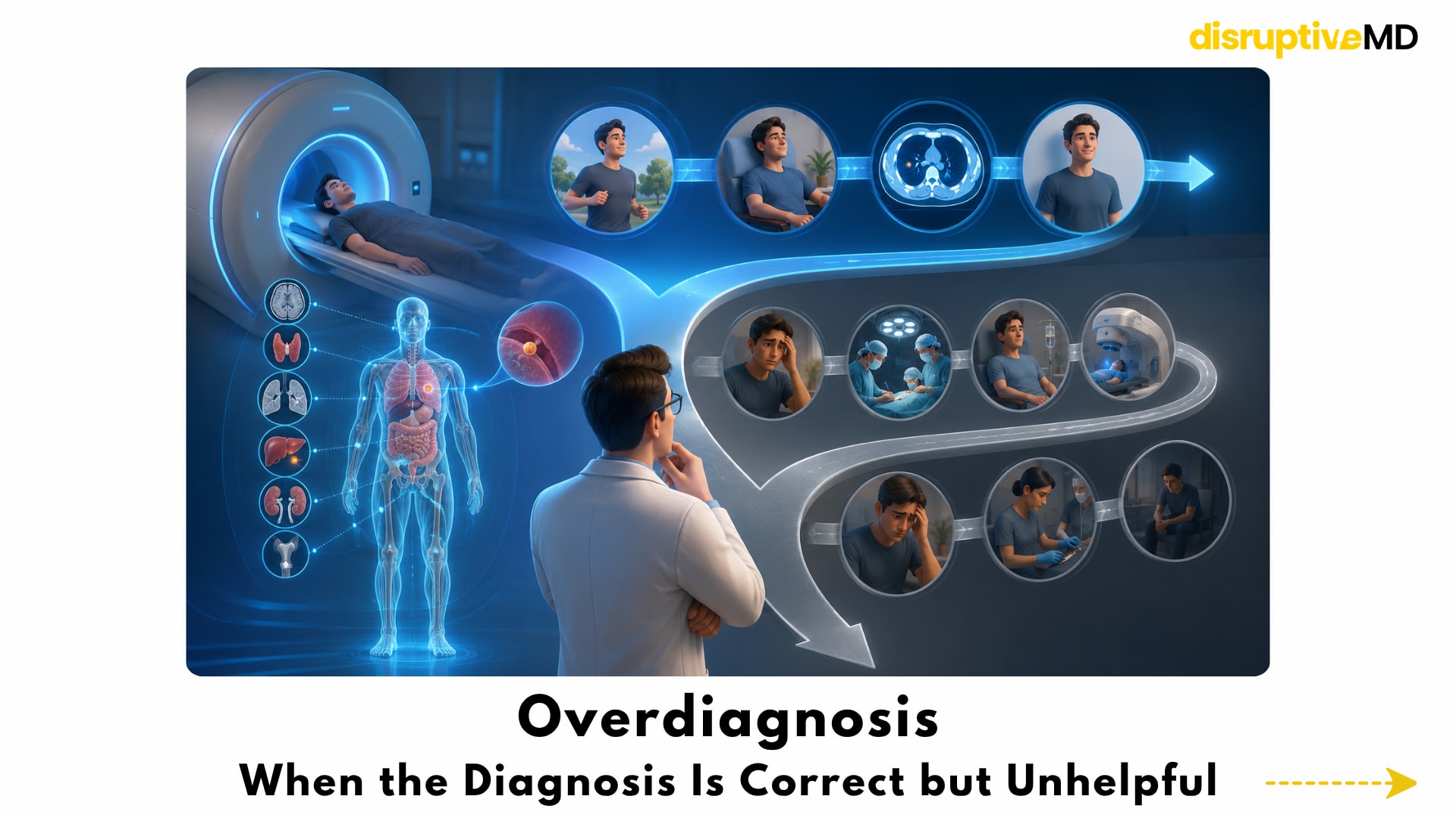
Overdiagnosis: When the Diagnosis Is Correct but Unhelpful
Overdiagnosis is different from a false positive.
In a false positive, the suspected disease is not actually present.
In overdiagnosis, the abnormality or disease is real—but it would never have caused symptoms, disability or death during the person’s lifetime.
This may occur when screening detects:
- an extremely slow-growing tumour;
- a biologically inactive cancer;
- a lesion that would never progress;
- a disease that would have remained clinically silent;
- an abnormality in a person who would die from another cause before it became harmful.
The diagnosis is technically accurate.
The problem is that the person cannot be helped by knowing about it.
Once detected, however, the disease may be treated.
This converts overdiagnosis into overtreatment.
Possible consequences include surgery, radiation, medication, organ damage, bleeding, infection, pain, reduced function and long-term psychological effects.
Overdiagnosis has long been recognised as one of the fundamental harms of screening. It occurs because many diseases exist across a spectrum, from rapidly progressive to biologically insignificant, and imaging cannot always predict their future behaviour at the time of detection.
This is one of the most difficult ideas in preventive medicine: It is possible to find a real disease, treat it successfully and still not have improved the patient’s life.
Why Cancer Detection Rate Is Not Enough
The most recent systematic review and meta-analysis included ten studies and 9,024 asymptomatic participants. The pooled rate of confirmed cancer detection was 1.57%.
That figure may sound impressive. It means that potentially important cancers were discovered in a small but meaningful proportion of participants.
However, most of the included studies had a moderate-to-serious risk of bias. Protocols were not standardised, incidental findings were frequent, and long-term outcome and cost-effectiveness data were lacking.
A detection rate cannot answer several essential questions:
- Would the cancers have become symptomatic later?
- Were they biologically aggressive?
- Did earlier detection change treatment?
- Did screening reduce advanced cancer?
- Did it reduce cancer-related mortality?
- How many benign abnormalities were investigated?
- How many invasive procedures were unnecessary?
- How many participants experienced lasting anxiety?
- How often did the scan miss disease?
- Was the programme cost-effective?
A screening programme should ultimately be evaluated by meaningful health outcomes, and not simply by the number of abnormalities found.
Lead-Time Bias
Full-body screening can also create the appearance of longer survival without changing the time of death.
Imagine that a cancer would normally become symptomatic at age 65 and cause death at age 70.
Without screening, the person appears to survive five years after diagnosis.
If a scan finds the same cancer at age 60 but treatment does not change its course, the person appears to survive ten years after diagnosis.
Survival after diagnosis has doubled.
But the person still dies at age 70.
The scan has increased the measured survival time without extending life.
This is known as lead-time bias.
It is one reason why five-year survival after diagnosis is not enough to prove that a screening test saves lives.
Length-Time Bias
Screening is also more likely to detect slowly growing disease than rapidly growing disease.
A slow-growing tumour remains in a detectable, symptom-free phase for longer. It therefore has a greater chance of being found during a scheduled scan.
An aggressive tumour may arise and become symptomatic between two screening examinations.
This means screening programmes can appear highly successful because they preferentially detect diseases that already have a more favourable biology.
The programme may report many early-stage cancers and excellent survival, even if its effect on aggressive disease is limited.
This is known as length-time bias.
It reinforces a crucial point: The diseases most easily detected through repeated screening may not always be the diseases most likely to kill.
The Peace-of-Mind Illusion
Many people purchase full-body scans because they want reassurance.
The emotional promise is understandable:
“I have checked everything. Nothing serious is hiding inside me.”
But no scan can examine every disease process with complete accuracy.
A normal result does not guarantee:
- that no cancer is present;
- that microscopic disease does not exist;
- that disease will not develop soon afterwards;
- that every organ was examined with the ideal protocol;
- that the scan replaced other screening tests;
- that new symptoms can safely be ignored.
Full-body MRI has recognised blind spots. Some areas are better assessed through dedicated imaging, laboratory testing, endoscopy, physical examination or other specialised tests.
A normal scan may therefore create false reassurance.
The opposite is also true.
An abnormal result does not necessarily mean that a serious disease is present.
Full-body screening can produce both unnecessary fear and excessive confidence.
True reassurance does not come simply from receiving a long imaging report. It comes from understanding what the examination can detect, what it may miss and what the findings actually mean.
One Scan Cannot Replace Established Screening
A full-body MRI is broad, but it is not a universal replacement for targeted screening.
Different diseases require different approaches.
A broad scan may not reproduce the sensitivity, resolution or evidence base of tests specifically designed for particular organs.
Depending on the individual, targeted screening may include:
- breast imaging;
- cervical screening;
- colorectal screening;
- low-dose lung imaging in selected high-risk people;
- blood-pressure assessment;
- lipid and glucose testing;
- skin examination;
- bone-health assessment;
- genetic counselling;
- organ-specific ultrasound;
- endoscopic examinations.
Whole-body imaging should not create the impression that every other preventive measure has become unnecessary.
The futuristic goal should not be one scan that replaces medicine.
It should be a coordinated system in which the right test is used for the right risk.
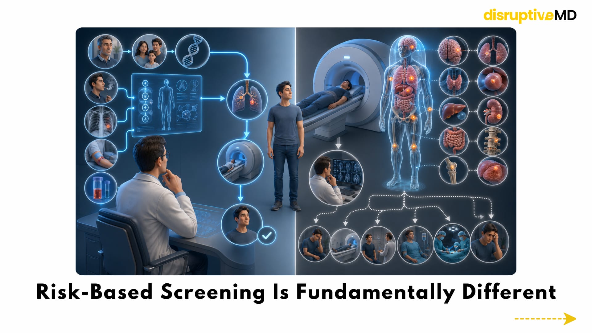
Risk-Based Screening Is Fundamentally Different
Established screening programmes generally begin with a defined risk.
The person may be selected based on: age;
- smoking exposure;
- symptoms;
- family history;
- inherited genetic variants;
- previous disease;
- occupational exposure;
- abnormal laboratory results;
- a known precancerous condition.
The screening test is then chosen to answer a specific clinical question.
Full-body screening reverses this process.
It looks across many organs first and asks which findings matter afterwards.
That approach can occasionally reveal a serious hidden disease.
It can also produce a large number of low-probability abnormalities in people who were unlikely to have the disease in the first place.
When disease prevalence is low, even a technically strong test can produce a meaningful number of false alarms.
Risk selection improves the probability that a positive finding represents something clinically important.
This may ultimately be the most important difference between useful surveillance and indiscriminate scanning.
When Whole-Body Imaging Makes More Sense
Whole-body MRI has stronger clinical justification in selected high-risk populations.
People with certain inherited cancer-predisposition syndromes can develop multiple cancer types in different organs, sometimes at young ages.
For these individuals, scanning only one organ may not be sufficient.
A meta-analysis involving 578 people with disease-causing TP53 variants found that baseline whole-body MRI identified new localised primary cancers in approximately 7% of participants. Most of the detected cancers were treated with curative intent.
A newer updated meta-analysis has continued to support the ability of whole-body MRI to detect cancers in asymptomatic people with this inherited high-risk condition.
Whole-body MRI also has established or developing roles in:
- assessing the extent of certain known cancers;
- evaluating bone-marrow disease;
- detecting multiple skeletal lesions;
- monitoring selected systemic diseases;
- surveillance for particular inherited conditions;
- evaluating patients with defined clinical indications.
These uses should not be confused with routine screening of every healthy adult.
The same scan can have a very different benefit-to-harm balance depending on who receives it.
High Risk and Low Risk Are Not the Same Population
Consider two individuals.
The first has a disease-causing genetic variant associated with a very high lifetime risk of multiple cancers.
The second is a young, asymptomatic person with no significant family history or known risk factors.
The scanner may be identical.
The medical decision is not.
In the high-risk patient, the chance of finding meaningful disease is substantially greater. Repeated surveillance may be justified despite the possibility of false positives.
In the low-risk individual, serious disease is less common. A larger proportion of detected abnormalities may therefore be benign, uncertain or clinically insignificant.
Screening must always be interpreted in the context of the probability of disease before the scan begins.
Commercial Wellness Scanning
Full-body MRI has moved beyond specialist surveillance and into consumer wellness.
The attraction is easy to understand.
The service offers:
- access without waiting for symptoms;
- a sense of control;
- advanced technology;
- a detailed report;
- the possibility of early detection;
- an experience that feels personalised and proactive.
Occasionally, these scans identify serious disease at an early stage.
Those stories are real and medically important.
But individual success stories cannot establish whether routine scanning improves outcomes across an entire healthy population.
For every dramatic early diagnosis, a complete evaluation must also count:
- benign findings;
- repeat scans;
- biopsies;
- surgeries;
- missed disease;
- overdiagnosis;
- anxiety;
- cost;
- false reassurance.
The central issue is not whether a commercial scan can ever find something important. The issue is whether the benefits are predictable, reproducible and greater than the harms for the population being offered the scan.
At present, the long-term answer remains uncertain.
The Reporting Problem
When a scan examines many organs, consistent reporting becomes essential.
Without a standardised system, two readers may classify the same abnormality differently. One may recommend immediate investigation, while another may recommend observation.
Structured reporting systems are being developed to classify whole-body MRI findings according to estimated cancer risk and to guide follow-up.
A five-category framework evaluated in 2,064 asymptomatic participants showed that higher imaging categories were associated with a higher probability of confirmed cancer. Confirmed cancer was present in 1.2% of the full cohort, while approximately half of the findings placed in the higher-suspicion categories were ultimately malignant.
This is an important step.
Better classification may reduce unnecessary alarm and create more consistent management.
But classification does not prove that population-wide screening improves survival.
It improves the handling of findings after the scan. It does not, by itself, prove that performing the scan was beneficial.
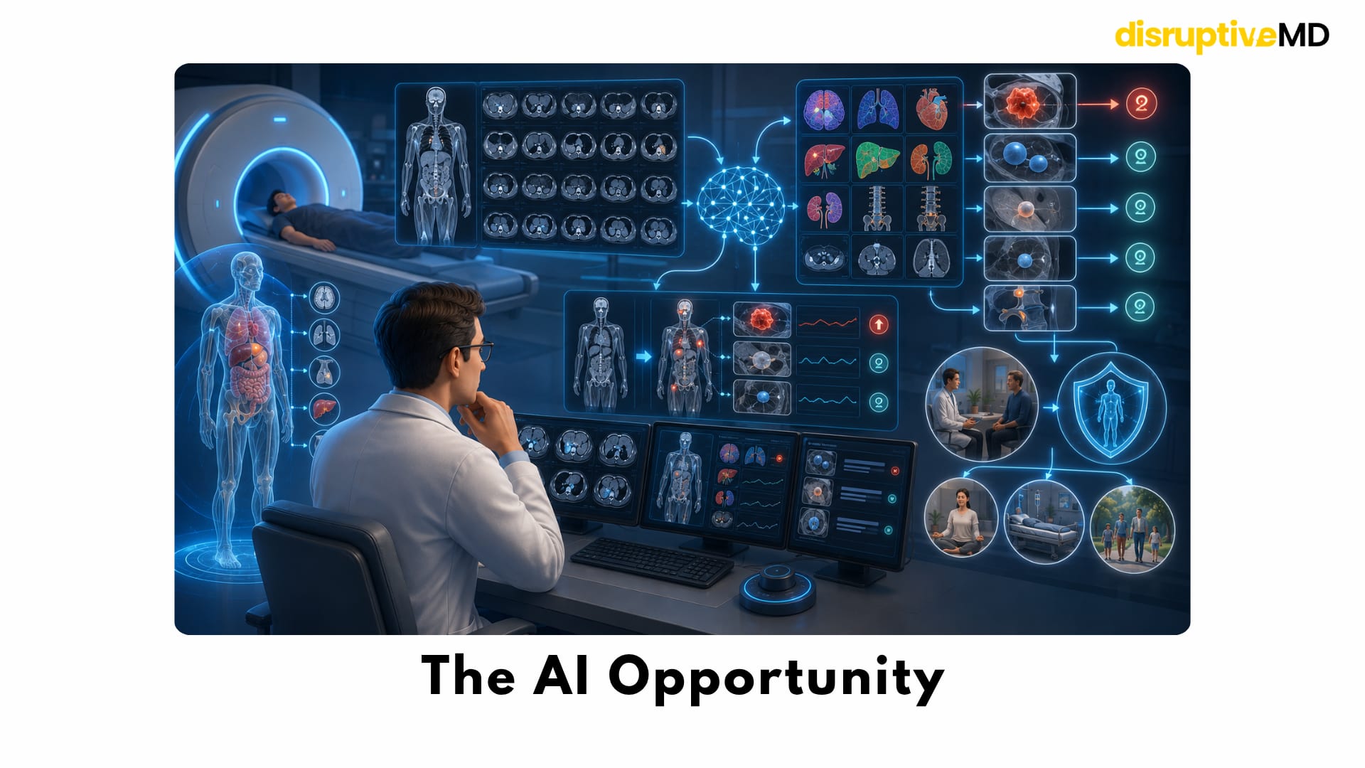
The AI Opportunity
Artificial intelligence could reshape full-body imaging.
A single whole-body examination may contain thousands of images. Reviewing every organ, tissue compartment and anatomical region is demanding and time-consuming.
AI may eventually assist with:
- automated organ segmentation;
- lesion detection;
- measurement of abnormalities;
- comparison with previous scans;
- identifying interval change;
- risk scoring;
- prioritising suspicious examinations;
- generating structured reports;
- recommending evidence-based follow-up;
- reducing variation between readers;
- shortening scan acquisition and reconstruction time.
The most valuable AI system would not simply detect more abnormalities.
It would help distinguish:
- aggressive from indolent disease;
- dangerous lesions from harmless variants;
- abnormalities requiring biopsy from those safe to observe;
- meaningful change from normal biological variation.
That is a much harder problem.
An algorithm trained to maximise sensitivity may identify more subtle abnormalities but also generate more false positives.
An algorithm trained to reduce false positives may miss early disease.
The future depends on finding the correct balance.
AI should therefore be judged by patient outcomes, not merely image-recognition accuracy.
AI Cannot Solve the Biology Problem Alone
A scan captures appearance.
Disease behaviour depends on biology.
Two lesions of similar size and shape may have very different molecular characteristics. One may remain stable for years. The other may progress rapidly.
Imaging-based AI can identify patterns that humans may not see, but its predictions must be validated across different scanners, populations and clinical settings.
It must also be tested for:
- false reassurance;
- hidden bias;
- performance across age groups;
- performance across sexes;
- variation between ethnic populations;
- rare diseases;
- unusual anatomy;
- differences in scan protocols;
- changes in performance over time.
Most importantly, the system must show that its recommendations improve outcomes without creating excessive follow-up.
A highly accurate algorithm can still be clinically harmful if it identifies conditions that do not need treatment.
Multi-Omics and Imaging
The most credible future may not involve scanning everyone.
It may involve deciding who should be scanned using a combination of biological and clinical data.
Future risk models may integrate:
- family history;
- inherited genetic variants;
- blood biomarkers;
- circulating tumour signals;
- metabolic measurements;
- proteomic patterns;
- immune markers;
- microbiome information;
- wearable trends;
- sleep and activity data;
- environmental exposure;
- previous imaging;
- age and clinical history.
Imaging could then be used selectively in people whose combined risk exceeds a meaningful threshold.
This would move preventive medicine away from: “Scan everyone and investigate whatever appears.”
And toward:“Estimate risk first, then select the examination most likely to help.”
Early research has already explored combining whole-genome sequencing, metabolomics, advanced imaging and clinical assessment. These studies show the technical possibility of building deeply personalised health profiles, but they also reveal large numbers of findings and the need for careful interpretation.
More data do not automatically create better decisions.
The future system must reduce uncertainty rather than simply produce more of it.
Liquid Biopsy and Imaging
Blood-based multi-cancer detection is another rapidly developing area.
Instead of searching anatomically for a visible mass, these tests look for tumour-associated signals in the blood.
Theoretically, blood testing and imaging could complement each other.
A future pathway might involve:
- risk assessment based on age, history and genetics;
- blood testing for abnormal molecular signals;
- targeted or whole-body imaging to locate the suspected disease;
- AI-supported risk classification;
- confirmation using dedicated imaging or tissue analysis.
This could reduce the need to scan every person indiscriminately.
However, blood-based detection faces many of the same unresolved questions as full-body imaging:
- false positives;
- false negatives;
- overdiagnosis;
- uncertain tissue of origin;
- investigation of very small signals;
- proof of mortality reduction;
- cost-effectiveness.
Combining two powerful technologies does not automatically remove their limitations.
It may also combine their false alarms.
The Psychological Cost of Knowing
Preventive screening is not only a technical process.
It is an emotional intervention.
An uncertain finding can change how a healthy person views their body.
They may begin to feel medically fragile.
Normal sensations can become alarming. Every ache may be connected to the scan result. Follow-up appointments may dominate months of life.
Research examining psychological outcomes after whole-body MRI has found that abnormal findings can create distress, particularly during periods of uncertainty and follow-up. Long-term effects vary between individuals and may be influenced by personality, perceived risk and the way results are communicated.
Some people may feel reassured by additional information.
Others may become trapped in repeated surveillance.
A responsible screening programme should therefore include:
- informed consent before scanning;
- clear explanation of incidental findings;
- realistic discussion of false positives;
- a defined follow-up pathway;
- access to clinical interpretation;
- support for uncertain or indeterminate results.
Selling the scan without explaining the possible consequences is not informed preventive medicine.
Cost and Inequality
Full-body scans are expensive and are frequently purchased privately.
But the price of the initial scan is only one part of the economic impact.
Additional costs may include:
- specialist consultations;
- dedicated imaging;
- repeated surveillance;
- laboratory testing;
- biopsies;
- anaesthesia;
- surgery;
- treatment of complications;
- time away from work.
These costs may be paid by the individual, an insurer or the wider healthcare system.
There is also an opportunity cost.
MRI machines, radiologists, technologists and specialist appointments are limited resources. Expanding low-risk screening could compete with imaging for people who have symptoms, known disease or established high-risk indications.
At the same time, access may be concentrated among wealthier individuals.
This creates a strange inequality.
Affluent populations may receive more early detection—but also more overdiagnosis, more surveillance and more unnecessary intervention.
The ethical question is not simply whether everyone should have access to more scanning.
It is whether the scanning provides enough health benefit to justify its cost and resource use.
What Would Prove That Full-Body Screening Works?
To establish routine full-body scanning as an effective preventive strategy, research would need to move beyond detection studies.
Large, long-term trials would need to examine:
- reduction in advanced-stage cancer;
- reduction in disease-specific mortality;
- reduction in overall mortality;
- quality of life;
- false-positive rates;
- false-negative rates;
- biopsy rates;
- procedural complications;
- overdiagnosis;
- psychological effects;
- cost-effectiveness;
- differences between risk groups;
- the ideal age to begin;
- the ideal screening interval;
- the best imaging protocol;
- the value added beyond established screening.
These studies are difficult.
They require large populations and many years of follow-up because deaths are less common than abnormal findings.
But without this evidence, medicine risks confusing technological capability with clinical benefit.
Fact Base
What recent research shows
A 2026 systematic review and meta-analysis evaluated ten studies involving 9,024 asymptomatic participants. The pooled confirmed cancer-detection rate was 1.57%. However, study protocols varied, most studies had a moderate-to-serious risk of bias, and long-term outcome and cost-effectiveness data were lacking.
A previous systematic review of whole-body MRI in asymptomatic people found that critical or indeterminate incidental findings were substantially more common than confirmed serious disease. Verification and long-term follow-up were incomplete in many studies.
A 2024 retrospective study of 2,064 asymptomatic participants found confirmed cancer in 24 people, representing 1.2% of the cohort. Forty-three people had findings classified in the two highest suspicion categories, but several high-suspicion findings were benign and supplementary tests identified cancers that the broad MRI examination had not classified as highly suspicious.
A population-based cohort study found that reporting tumour-related incidental findings increased biopsy activity. Some biopsies detected malignancies, while many identified benign disease.
Research in inherited high-risk populations has shown a higher cancer yield than that seen in average-risk screening. In one meta-analysis of people with disease-causing TP53 variants, baseline whole-body MRI detected new localised primary cancers in approximately 7% of participants.
MRI does not involve ionising radiation, making it more suitable than CT for repeated imaging when whole-body surveillance is clinically justified. CT is fast and highly effective for several anatomical regions, but it uses ionising radiation. In asymptomatic people, particularly when examinations are repeated, this exposure must be justified by a meaningful expected benefit.
Overdiagnosis is a genuine cancer-screening harm. It involves correctly detecting a cancer that would never have caused symptoms or death, potentially leading to treatment from which the patient could not benefit.
No single whole-body protocol performs equally well for every organ and every disease.
Detection of cancer is not the same as proof that screening reduces mortality.
What the evidence does not yet establish
Current research has not established that routine full-body MRI helps healthy, low-risk adults live longer.
The ideal starting age and repeat interval for average-risk screening remain unknown.
The balance between cancers detected and people harmed through unnecessary follow-up remains uncertain.
The long-term rate of overdiagnosis is not yet known.
Cost-effectiveness has not been established for broad population screening.
AI has not yet eliminated false positives, false negatives or overdiagnosis.
Multi-omics and imaging integration remains promising but experimental as a population-wide screening pathway.
What Is Real Today
Full-body imaging can reveal hidden abnormalities before symptoms appear.
Some clinically important cancers and vascular abnormalities will occasionally be found early.
MRI can examine large areas of the body without ionising radiation.
CT offers speed and strong visualisation of several anatomical structures but involves radiation exposure.
Incidental findings are common.
Many detected abnormalities are benign or uncertain.
False positives can lead to additional scans, biopsies, procedures, anxiety and cost.
Whole-body MRI has a stronger role in selected high-risk populations than in healthy, low-risk adults.
Structured reporting can improve consistency and guide follow-up.
Full-body imaging cannot replace every established organ-specific screening test.
What Is Not Fully Real Yet
A scan that reliably separates every dangerous disease from every harmless finding does not exist.
There is no proof that routine full-body scanning increases lifespan in healthy, low-risk adults.
AI cannot yet predict the future behaviour of every detected lesion.
A normal scan cannot guarantee future health.
A single scan cannot replace clinical assessment, family history, risk evaluation, laboratory testing or established screening.
The best interval for repeating full-body scans in average-risk adults is unknown.
A universal scan-and-treat preventive model has not been validated.
A fully integrated system combining imaging, genetics, biomarkers, wearables and AI remains a future vision rather than routine clinical reality.
Key Takeaway
Full-body scanning sits at the edge of preventive medicine and medical overreach.
It represents both the promise and the danger of modern diagnostics. The promise is clear:
A serious disease could be detected before symptoms begin, while treatment is still possible.
The danger is equally clear:
The scan may identify harmless abnormalities, create anxiety, trigger unnecessary procedures and diagnose diseases that never needed to be found.
The central challenge is no longer simply seeing inside the body.
It is understanding what deserves attention.
The future of preventive medicine may not be scanning every healthy person as often as possible.
It may be using genetics, biomarkers, clinical history, environmental exposure and AI to determine exactly who is likely to benefit—followed by imaging designed to answer a specific question.
**We are becoming extremely good at finding things inside the human body. The next medical breakthrough will be knowing which findings truly matter, and which ones should be left alone. **
References
- Hu YS, Wu CA, Lin DC, et al. Applying ONCO-RADS to whole-body MRI cancer screening in a retrospective cohort of asymptomatic individuals. Cancer Imaging. 2024;24:22. DOI: https://doi.org/10.1186/s40644-024-00665-z
- Brenner DJ, Elliston CD. Estimated radiation risks potentially associated with full-body CT screening. Radiology. 2004;232(3):735–738. DOI: https://doi.org/10.1148/radiol.2323031095
- Kwee RM, Kwee TC. Whole-body MRI for preventive health screening: a systematic review of the literature. Journal of Magnetic Resonance Imaging. 2019;50(5):1489–1503. DOI: https://doi.org/10.1002/jmri.26736
- Gibson LM, Paul L, Chappell FM, et al. Potentially serious incidental findings on brain and body magnetic resonance imaging of apparently asymptomatic adults: systematic review and meta-analysis. BMJ. 2018;363:k4577. DOI: https://doi.org/10.1136/bmj.k4577
- Richter A, Sierocinski E, Singer S, et al. The effects of incidental findings from whole-body MRI on the frequency of biopsies and detected malignancies or benign conditions in a general population cohort study. European Journal of Epidemiology. 2020;35(10):925–935. DOI: https://doi.org/10.1007/s10654-020-00679-4
- Welch HG, Black WC. Overdiagnosis in cancer. Journal of the National Cancer Institute. 2010;102(9):605–613. DOI: https://doi.org/10.1093/jnci/djq099
- Martins da Fonseca J, Trennepohl T, Pinheiro LG, et al. Whole-body MRI for opportunistic cancer detection in asymptomatic individuals: a systematic review and meta-analysis. European Radiology. 2026;36(3):1813–1823. DOI: https://doi.org/10.1007/s00330-025-11976-5
- Ballinger ML, Best A, Mai PL, et al. Baseline surveillance in Li-Fraumeni syndrome using whole-body magnetic resonance imaging: a meta-analysis. JAMA Oncology. 2017;3(12):1634–1639. DOI: https://doi.org/10.1001/jamaoncol.2017.1968
- Dacoregio MI, Abrahão Reis PC, Gonçalves Celso DS, et al. Baseline surveillance in Li-Fraumeni syndrome using whole-body MRI: a systematic review and updated meta-analysis. European Radiology. 2025. DOI: https://doi.org/10.1007/s00330-024-10983-2
- Perkins BA, Caskey CT, Brar P, et al. Precision medicine screening using whole-genome sequencing and advanced imaging to identify disease risk in adults. Proceedings of the National Academy of Sciences. 2018;115(14):3686–3691. DOI: https://doi.org/10.1073/pnas.1706096114
- Hou YC, Yu HC, Martin R, et al. Precision medicine integrating whole-genome sequencing, comprehensive metabolomics, and advanced imaging. Proceedings of the National Academy of Sciences. 2020;117(6):3053–3062. DOI: https://doi.org/10.1073/pnas.1909378117
- Conti L, Mazzoni D, Marzorati C, et al. Observations regarding the detection of abnormal findings following a cancer-screening whole-body MRI in asymptomatic subjects: the psychological consequences and the role of personality traits over time. Journal of Magnetic Resonance Imaging. 2025;61(2):634–645. DOI: https://doi.org/10.1002/jmri.29461
- Petralia G, Koh DM, Attariwala R, et al. Oncologically relevant findings reporting and data system: guidelines for the acquisition, interpretation, and reporting of whole-body MRI for cancer screening. Radiology. 2021;299(3):494–507. DOI: https://doi.org/10.1148/radiol.2021201740
- Basar Y, Alis D, Tekcan Sanli DE, Akbas T, Karaarslan E. Whole-body MRI for preventive health screening: management strategies and clinical implications. European Journal of Radiology. 2021;137:109584. DOI: https://doi.org/10.1016/j.ejrad.2021.109584
- Cieszanowski A, Maj E, Kulisiewicz P, et al. Non-contrast-enhanced whole-body magnetic resonance imaging in the general population: the incidence of abnormal findings in patients 50 years old and younger compared with older subjects. PLoS ONE. 2014;9(9):e107840. DOI: https://doi.org/10.1371/journal.pone.0107840
- Hegenscheid K, Seipel R, Schmidt CO, et al. Potentially relevant incidental findings on research whole-body MRI in the general adult population: frequencies and management. European Radiology. 2013;23(3):816–826. DOI: https://doi.org/10.1007/s00330-012-2636-6