
Introduction
Artificial intelligence is transforming radiology, but the most precise issue is not will AI replace radiologists. The better question is what parts of radiology will be automated, what parts will be AI-assisted, and what areas will still need physician judgment. While radiology is sometimes regarded as an image-reading field, clinical radiologists do far more than spot patterns in x-rays, CT scans, MRIs, ultrasounds and mammograms. They connect imaging findings with symptoms, lab results, prior scans, cancer history, surgical plans, emergency priority, procedural decisions, and communication with treating teams. Existing data suggest that AI can improve certain imaging operations like detection, triage, measurement, reconstruction and workflow prioritization, but is not yet justified in replacing radiologists as responsible clinical decision-makers.
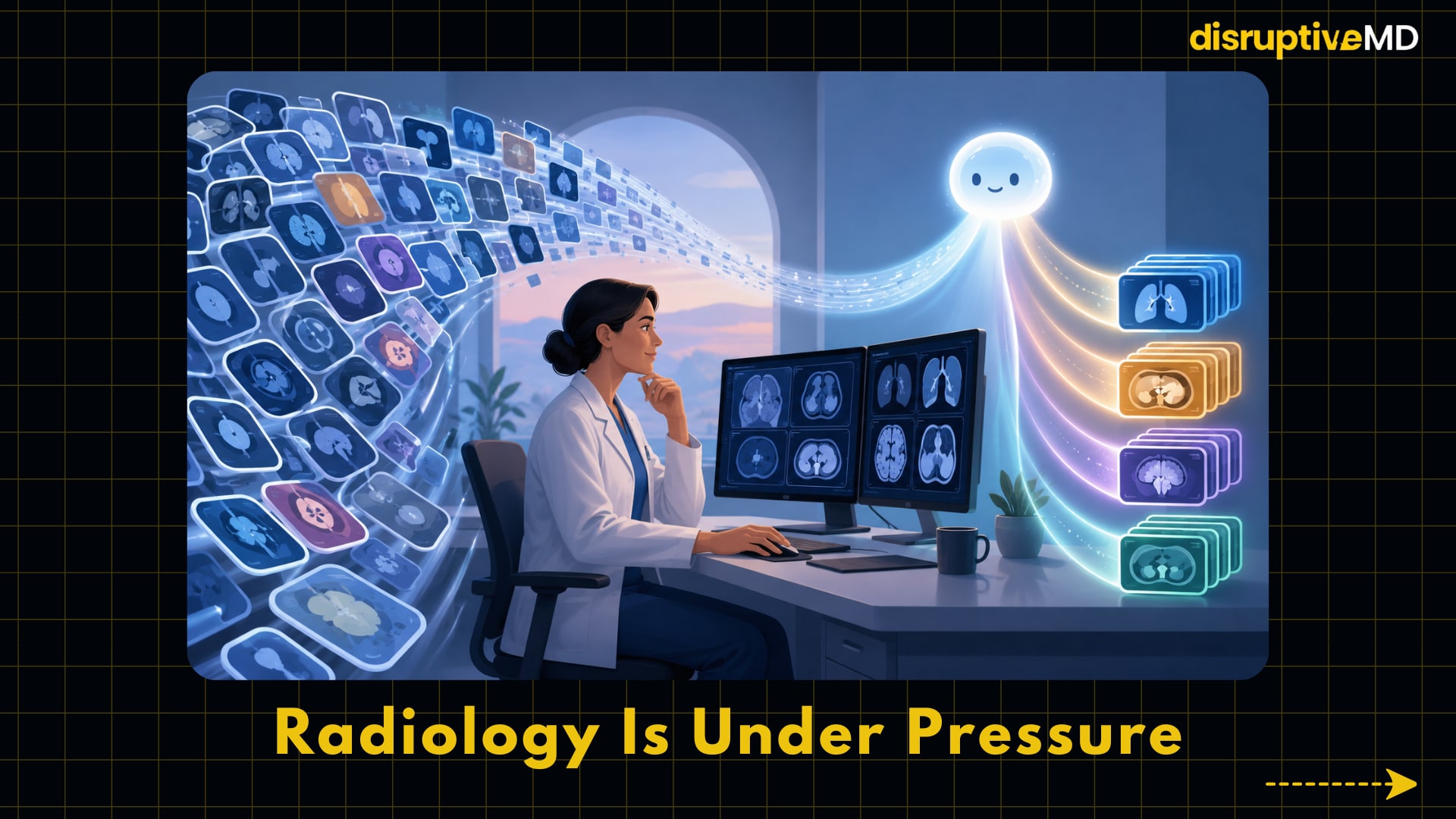
The Real Background: Radiology Is Under Pressure
The challenge in radiology is load. Imaging is essential to modern health care, including diagnosis, treatment planning, emergency care, cancer staging, surgical navigation and follow-up. A 2025 RSNA special report in the United States described a shortage of radiologists where the demand for imaging outpaces the growth of the radiology workforce . The report also noted that the average workload of radiologists nearly doubled from 14,900 studies/year in 2008 to 26,457 studies/year in 2018, while the number of radiologists entering the workforce increased by only 13% .This is significant because delays in interpretation of imaging can lead to delays in diagnosis, decisions on therapy, release from emergency departments and cancer care. Artificial intelligence in radiology is usually related with machine learning software that learns statistical patterns from medical images and associated data. A frequent type is deep learning, which employs layered artificial neural networks to identify patterns like lung nodules, intracranial bleeding, breast masses, fractures, organ edges, or image quality features. They don't "understand" sickness the way clinicians do, they compute likelihood based on patterns in data. That distinction is crucial, since a high probability imaging discovery still needs to be examined in a clinical context of the patient.


- AI is transforming radiology, but the real question is which tasks can be automated and which still require physician judgment.
- Radiologists do more than read images; they connect scans with symptoms, labs, prior imaging, cancer history, surgical plans, and clinical urgency.
- Current AI can assist detection, triage, measurement, reconstruction, and workflow prioritization, but it is not ready to replace radiologists as clinical decision-makers.
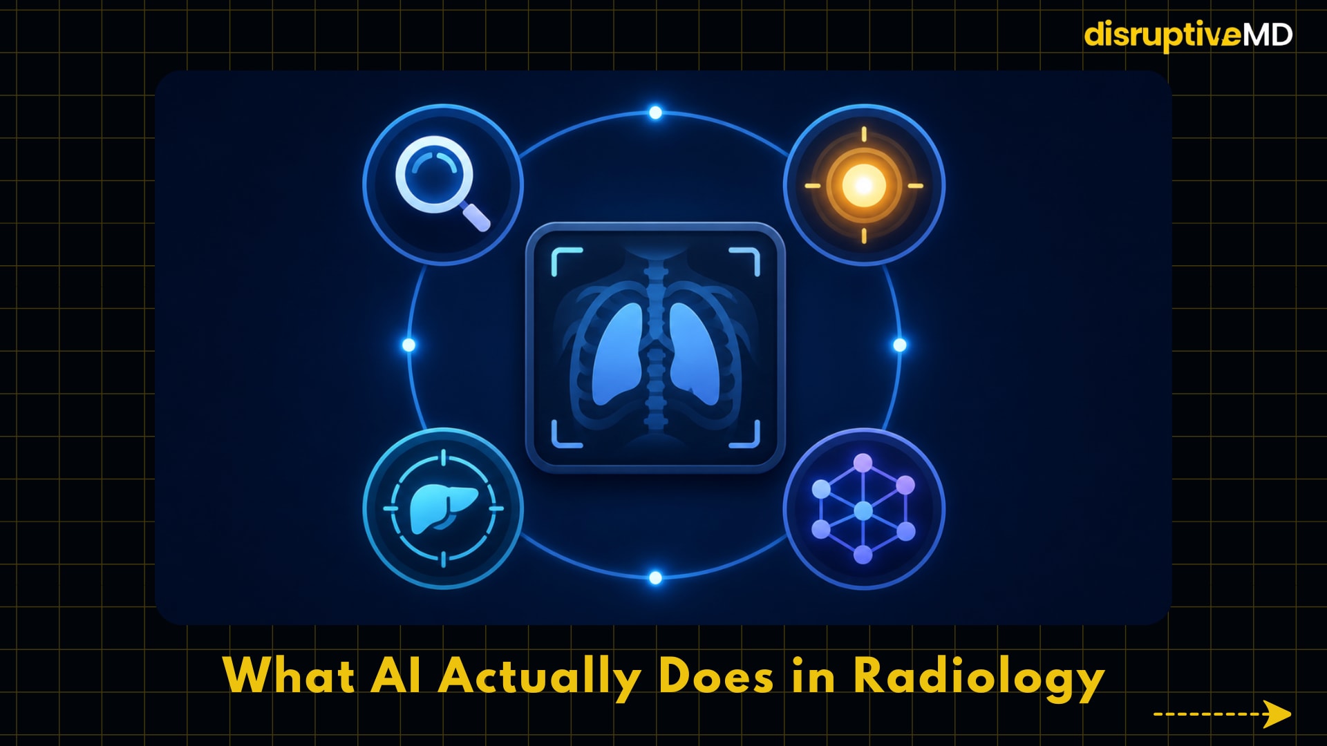
What AI Actually Does in Radiology
Most of the AI solutions for radiology are created today to do particular jobs, not to do the whole job of a radiologist. CADe (computer-aided detection) spots suspicious regions, such as possible lung nodules or breast cancers. Computer assisted diagnosis (CADx) estimates the probability that a finding is a given disease. Triage software can assist in prioritizing urgent studies, such as a suspected stroke or brain bleed, so radiologists can review those sooner. Quantification tools measure structures like tumor volume, heart chamber size, bone density or organ boundaries. These services can be useful in radiology because there are many repetitive, time-sensitive, measurement-heavy occupations where ongoing help might decrease cognitive load. The list of AI-enabled medical devices by the U.S. FDA indicates the rapid expansion in this area, but also proves that regulatory clearance does not imply total clinical substitution. According to the FDA, the list of listed devices has met the applicable premarket standards, including the focused examination of safety and effectiveness for their intended use. The FDA also warns that the list is not comprehensive and public summaries do not contain all the material submitted. Analysis of 1,016 devices showed that 84.4% of unique devices used images as the primary input, and that radiology was the most common review panel for 88.2% of image-based devices, indicating radiology as the dominant clinical domain for medical AI deployment. Image inputs and radiography were the most popular aspects for FDA AI/ML-enabled medical device authorizations in 2025. In practical terms, this means that AI today is less like a totally independent physician, and more like a highly skilled assistant. It might identify a likely pulmonary embolism, delineate a tumour, assess risk of breast cancer, enhance an MRI reconstruction or reorder a worklist. But it is the radiologist who has the ultimate responsibility to judge whether the finding is real, of clinical relevance, fits the patient’s history and is properly communicated. This is crucial because accidental observations, artifacts, postoperative alterations, unusual diseases, and lack of clinical information are common in medical pictures and require judgment beyond a single algorithmic result.
Evidence and Real-World Meaning
Today, the clearest evidence for AI support in radiology is in very specialized and well defined use cases such as screening mammography. In the MASAI randomized controlled research, 105,934 women in Sweden were randomized to either AI-assisted mammography screening or standard double reading without AI. AI was used for triage of examinations to single or double reading by radiologists and for detection support. In the 2026 Lancet research, the risk of interval cancers was 1.55 per 1,000 people in the AI-assisted group and 1.76 per 1,000 people in the control group. AI group has higher sensitivity (80.5%) than control group (73.8%) but the same specificity (98.5%) . Sensitivity is the ability to correctly identify individuals who have disease, while specificity is the ability to correctly identify those who do not have disease. The MASAI findings are noteworthy because interval cancers are those diagnosed between one negative screening test and the next scheduled screening round. These malignancies are clinically significant as they are often missed or represent fast advancing illness. In the AI-supported group, the trial showed a lower number of interval cancers with adverse features. Previous safety studies of MASAI showed a 44% reduction in screen-reading burden and in a later investigation a 29% increase in cancer diagnosis with no increase in false positive. False positive: A test result that shows cancer when there is no cancer. That can mean further imaging, biopsies, stress and money. Real-world deployments also provide evidence to support the premise that AI can improve certain screening procedures. A nationwide German mammography study of 463,094 women screened, including 260,739 women screened with AI support, found that the breast cancer detection rate was 6.7 per 1,000 in the AI-supported group vs. 5.7 per 1,000 in the control group, representing a relative increase of 17.6%, without adversely affecting recall rate. Recall rate is the proportion of patients who are asked back for further testing after screening. Why does this matter? An AI system that detects more cancers but also substantially increases recalls potentially harm patients through over-testing. The German data suggests that in this situation detection went up without that trade-off. But the whole body of data across radiology is a little more mixed than the best mammography studies reveal. A quick systematic scoping review (January 2020-January 2025) of 140 papers in eClinicalMedicine found evidence of enhanced diagnostic accuracy and reduced interpretation time but data on user experience, false positives, workflow efficiency and cost-effectiveness was mixed. In 2024, a comprehensive review and meta-analysis of studies evaluating real-world imaging workflows found that while 67% of studies measuring task time reported decreases, pooled meta-analyses showed no meaningful impacts, largely due to large heterogeneity in study methodology and clinical circumstances . So the meaning in the real world is particular, not universal. AI can assist radiologists in working faster and may improve detection in specific validated tasks, however performance is dependent on disease, imaging modality, patient demographic, scanner type, workflow architecture and clinician response to the AI output. An algorithm that has been validated in a national screening program cannot be assumed to perform equally well in emergency CT, pediatric imaging, low-resource settings, rare cancers, post-surgical anatomy, or populations underrepresented in training data.
Limitations, Risks, and Unanswered Questions
The major limitation is clinical generalizability – can an AI tool that works well on one data set or at one hospital continue to perform safely across varied patients, scanners, sickness patterns, races, age groups, and healthcare systems. In 2025, an examination of 903 FDA-approved AI-enabled medical devices, published in JAMA Network Open, found that 692 (76.6%) were in radiology but only 55.9% had clinical performance studies, 8.1% had prospective nonrandomized studies, and 2.4% had randomized trials. Few clinical assessments reported results stratified by sex (less than one third) and only around one fourth reported results on subgroups defined by age. Bias is another significant problem, since AI systems learn from the data they are taught and evaluated on. The algorithm may be less reliable for certain populations, disease presentations, scanners, body types, or health care settings if the training data are not representative of those groups. In a 2024 npj Digital Medicine scoping investigation of FDA-approved AI/ML medical devices, just 3.6% of approvals identified race or ethnicity, 99.1% of approvals did not provide any socioeconomic data, and participant age was not reported in 81.6% of approvals . This matters because radiology AI could be generally accurate but bad for some groups of patients. Another real danger is automation bias Automation bias is when the physician trusts too much a recommendation made by a computer, especially when he is busy or he may miss something that the AI system does not point out . The other concern is also possible: If AI creates too many alerts or false positives, physicians may suffer from alert fatigue and ignore valuable messages. To enhance therapy, AI should be integrated into workflow in a manner that aids human judgment, not silently usurps it. In 2024, major radiology organizations issued a multi-society statement that emphasized practical, ethical and safety considerations such as tracking the utility and safety of AI after deployment. Cost and Access not resolved Artificial intelligence may reduce some reading effort in some workflows, but health organizations still need to license software, integrate technology, establish cybersecurity safeguards and monitoring systems, train staff, assure quality, comply with medicolegal regulations, and plan for algorithm improvements. Limited evidence exists regarding cost-effectiveness and benefits may vary between high-volume university centers, community hospitals, rural imaging networks and low-resource settings. This is significant, because a strategy that improves efficiency in one system can add complexity or cost to a different system.
Conclusion
AI is not likely to replace radiologists as a profession in the foreseeable clinical environment, but it is already beginning to replace or change specific activities in radiology. The best current paradigm is “radiologist plus validated AI, with monitoring,” not “AI versus radiologist.” In this model, AI is used for certain repetitive or time-sensitive tasks, with radiologists still responsible for clinical interpretation, managing uncertainty, communication, procedures, interdisciplinary choices, and patient safety. For patients it promises faster reporting, earlier detection in some illnesses and more dependable measurement. For clinicians, the benefit is greater workflow help and less delays. Artificial intelligence may help health systems cope with the increasing demand for imaging, but only if the technologies are adequately vetted, monitored, equitable and integrated. AI may not replace the future radiologist, but the profession of radiology will increasingly be shaped by radiologists who know how to utilize AI properly, challenge it intellectually, and remain accountable for patient care.
Evidence Rating
Mixed or limited evidence. There are regulatory approved AI technologies in radiology and there is substantial clinical trial evidence in specific sectors such as AI supported mammography screening with randomized data from the MASAI project. However, there is limited evidence for a broad replacement of radiologists. Many AI devices require explicit, prospective, diversified or randomised clinical validation. The most evidence-based conclusion is that AI can assist radiologists with particular activities, but cannot replace the full clinical role of radiologists.
Educational Disclaimer
The material in this article is not intended to be a substitute for professional medical advice, diagnosis, or treatment. Patients should consult with qualified healthcare professionals for imaging, diagnosis or medical care.
References
- U.S. Food and Drug Administration. Artificial Intelligence-Enabled Medical Devices.
- Singh, R., Bapna, M., Diab, A.R. et al. How AI is used in FDA-authorized medical devices: a taxonomy across 1,016 authorizations. npj Digit. Med. 8, 388 (2025). https://doi.org/10.1038/s41746-025-01800-1.
- Gommers J, Hernström V, Josefsson V, Sartor H, Schmidt D, Hjelmgren A, Larsson AM, Hofvind S, Andersson I, Rosso A, Hagberg O, Lång K. Interval cancer, sensitivity, and specificity comparing AI-supported mammography screening with standard double reading without AI in the MASAI study: a randomised, controlled, non-inferiority, single-blinded, population-based, screening-accuracy trial. Lancet. 2026 Jan 31;407(10527):505-514. doi: 10.1016/S0140-6736(25)02464-X. PMID: 41620232.
- Lawrence R, Dodsworth E, Massou E et al. Artificial intelligence for diagnostics in radiology practice: a rapid systematic scoping review, eClinicalMedicine, 2025; 83.
- Wenderott, K., Krups, J., Zaruchas, F. et al. Effects of artificial intelligence implementation on efficiency in medical imaging—a systematic literature review and meta-analysis. npj Digit. Med. 7, 265 (2024). https://doi.org/10.1038/s41746-024-01248-9.
- Eisemann, Nora ; Bunk, Stefan ; Mukama, Trasias et al. / Nationwide real-world implementation of AI for cancer detection in population-based mammography screening. In: Nature Medicine. 2025 ; Vol. 31, No. 3. pp. 917-924.
- Windecker D, Baj G, Shiri I, et al. Generalizability of FDA-Approved AI-Enabled Medical Devices for Clinical Use. JAMA Netw Open. 2025;8(4):e258052. doi:10.1001/jamanetworkopen.2025.8052.
- Muralidharan, V., Adewale, B.A., Huang, C.J. et al. A scoping review of reporting gaps in FDA-approved AI medical devices. npj Digit. Med. 7, 273 (2024). https://doi.org/10.1038/s41746-024-01270-x.
- ACR, CAR, ESR, RANZCR, RSNA. Developing, Purchasing, Implementing and Monitoring AI Tools in Radiology: Practical Considerations. Multi-society statement summarized by Elsevier/ACR. 2024.
- Afshari Mirak S, et al. The Growing Nationwide Radiologist Shortage. Radiology. 2025.